2329: AMTX-100, a Nuclear Transport Inhibitor, Attenuates Inflammatory Cytokine Production in vitro and Following UV Mediated Skin Inflammation in a Mouse Model of Cutaneous Lupus Erythematosus in vivo
University of Washington Seattle, WA, United States
Disclosure information not submitted.
Xizhang Sun1, Jie An1, Ting Wang1, Arpit Rathee1, Vernon Alvarez2, Matthew Gonda3, Christian Lood1 and Keith Elkon1, 1University of Washington, Seattle, WA, 2Amytrx Therapeutics Inc., Nashville, TN, 3Amytrx Therapeutics, Nashville, TN
Background/Purpose: Inflammatory stimuli induce transcription factors (TFs) such as NF-kB and interferon regulatory factors (IRFs). TFs are transported from cytosol to nucleus to activate genes encoding chemokines and cytokines. Nuclear transport of TFs is performed by importins that comprise a beta chain and different alpha chains that confer substrate specificity for TF nuclear localization sequences (NLS). The alpha-5-containing importin (Impα5) is of particular interest because it transports STAT1, NF-kB, and other stress responsive TFs containing a NLS. AMTX-100 is a 28 amino acid chimeric peptide containing a huFGF4 domain and NF-kB p50 NLS that function to facilitate leukocyte cell penetration and as a nuclear transport checkpoint inhibitor (NTCI), respectively. AMTX-100 is currently in a Phase 2b clinical trial to treat inflammation in eczema. Since Impα5 binds to the NLS of NF-kB p50 as well as STAT1 & 3, we examined the effects of AMTX-100 on inflammatory pathways and Type I interferons (IFN-I), in vitro and in vivo.
Methods: The human monocyte cell line, THP-1 was transfected with 0.1 ug dsDNA in the presence or absence of AMTX-100 and cytokine mRNA expression quantified by qPCR. Neutrophils from healthy volunteers were isolated by density gradient and exposed to either PMA, the ionophore A23187 or immune complexes (IC) containing the lupus antigen SmRNP. NET formation was quantified by release of DNA and MPO as determined by fluorimetry. Female C57BL/6 (B6) mice aged 8-12 weeks were exposed to a single dose of UV (500mJ/cm2) on the dorsal skin. Skin biopsies were obtained before and at 6 and 24 hr post UV exposure. Cytokine gene expression was determined by qPCR. Statistical significance between groups was determined by Student's t-test.
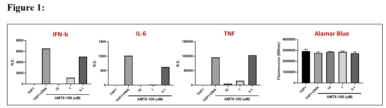
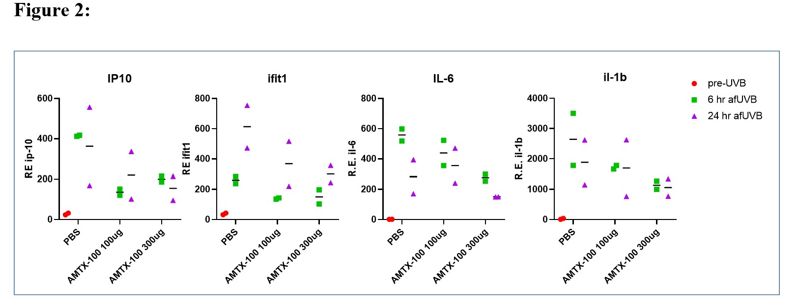
Results: In vitro studies with THP-1 cells revealed that 1 uM AMTX-100 inhibited IFN-b expression by 82% , and IL-6, TNF and IL-1b by 98%, 84%,and 95% respectively (Fig. 1). In neutrophils incubated with NET-inducing stimuli, AMTX-100 (3 uM) inhibited DNA and MPO release by ~40% in PMA or A23187 stimulated cells but did not affect NET release induced by IC. To determine whether AMTX-100 attenuated the IFN-I response following UV induced inflammation of mouse skin, we applied either 100 or 300 ug AMTX-100 in saline onto the skin before and once daily after UV exposure. As shown in Fig. 2, ISGs, as well IL-1 and IL6 were all reduced in an apparent dose dependent fashion. Similar results were obtained in a second in vivo experiment (not shown).
Conclusion: We conclude that AMTX-100 reduces Type I IFN and NF-kB-dependent inflammatory cytokines from DNA activated human monocytes in vitro. While AMTX-100 had a modest inhibitory effect on NET formation in response to PMA and ionophore stimulation, it did not attenuate NET formation induced by ICs in vitro. When tested in vivo using the lupus-relevant UV skin exposure, we observed a dose-dependent inhibition of several NF-kB-dependent cytokines as well as ISGs. Since AMTX-100 is in clinical trials for inflammatory skin disease, these findings suggest a novel therapeutic approach utilizing importin NTCIs that may be useful for prevention or treatment of cutaneous lupus erythematosus.
AMTX-100 inhibits IFN-b and inflammatory cytokine production in vitro. The THP-1 monocyte cell line was incubated with serial 10-fold dilution of AMTX-100 as shown on the X-axis. Left three panels: After 1 hr, cells were transfected with ds-DNA and 6 hrs later the cells were harvested and the RNA expression of IFN-b, IL-6 and TNF quantified by qPCR. The results are shown as expression of the cytokine relative to 18S RNA (R.E. on the Y-axis). Right Panel: The Alamar blue test for viability revealed that the AMTX-100 peptide was non-toxic at the doses used in these experiments.
AMTX-100 attenuates the skin IFN and inflammatory response to UV light exposure. C57BL/6 mice were exposed to a single dose of 500 mJ of UV light on the shaved dorsum. As shown on the X-axis, mice received either saline (PBS) or 100 or 300 ug AMTX-100 peptide in PBS before and after UV exposure. Biopsies were obtained at time 0 and at 6 and 24 hr after UV exposure. RNA was extracted and the relative expression of representative ISGs (IP10 and Ifit1) and the inflammatory cytokines (IL-6 and IL-1b) shown on the Y axis. Relative expression of genes prior to UV is shown in orange.
X. Sun: None; J. An: None; T. Wang: None; A. Rathee: None; V. Alvarez: Amytrx Therapeutics, 2; M. Gonda: Amytrx Therapeutics, Inc, 3, 4; C. Lood: Amytryx, 5, Boehringer-Ingelheim, 5, Bristol-Myers Squibb(BMS), 5, Citryll, 2, Eli Lilly, 5, Gilead, 5, Horizon Therapeutics, 5, Pfizer, 5, Redd Pharma, 5, 11; K. Elkon: None.