SUNY Upstate Medical University Syracuse, NY, United States
Disclosure information not submitted.
Christian Geier1, Haani Qudsi2, Jihad BenGabr1, Robert Winchester3 and Andras Perl4, 1SUNY Upstate Medical University, Syracuse, NY, 2Norton College of Medicine, SUNY Upstate Medical University, Syracuse, NY, 3Columbia University, New York, NY, 4SUNY, Syracuse, NY
Background/Purpose: A variant of HLA-DR confers the strongest genetic risk for rheumatoid arthritis (RA) suggesting that DRhi cells are important in RA. We previously found that RA blood contains 'unorthodox' DRhi immune cells (non-lymphoid cells that do not conform to bona fide definitions of monocytesnor dendritic cells).The purpose of the study was to determine whether DRhi immune cells expressing granulocyte associated molecules (CD15, CCR3)contribute to these alterations of the DRhi pool in RA.
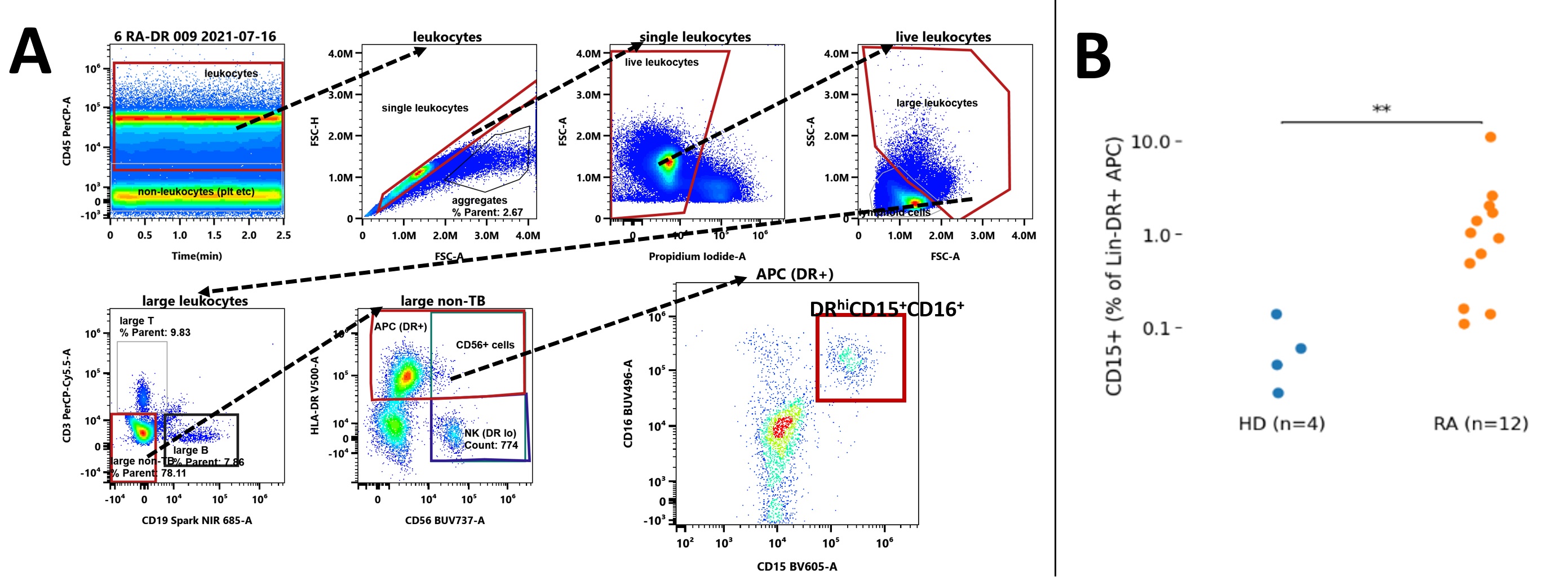
Methods: We studied RA patients (n=12) satisfying the 2010 ACR classification criteria and matched healthy donors by flow cytometry. PBMC and other low-density cells were isolated from blood by Ficoll density gradient centrifugation. To gate non-lymphoid DRhi cells we excluded lymphocytes and non-viable cells based onforward and side scatter, CD3/CD19 dump and viability gates (Fig. 1A). We quantified the contribution of CD15+ cells to the non-lymphoid DRhi pool and their expression (MFI) of co-stimulatory and co-inhibitory molecules. We used t-SNE on index patients (debilitating polyarticular synovitis)to clarify the higher-dimensional structure of the CD15positive DRhi subpopulation followed by bi-axial gating to validate anyt-SNE guided observation and quantify distinctive features of DRhiCD15+. Kruskal-Wallis testing with a threshold of p< 0.05 was performed to assess for significant differences.
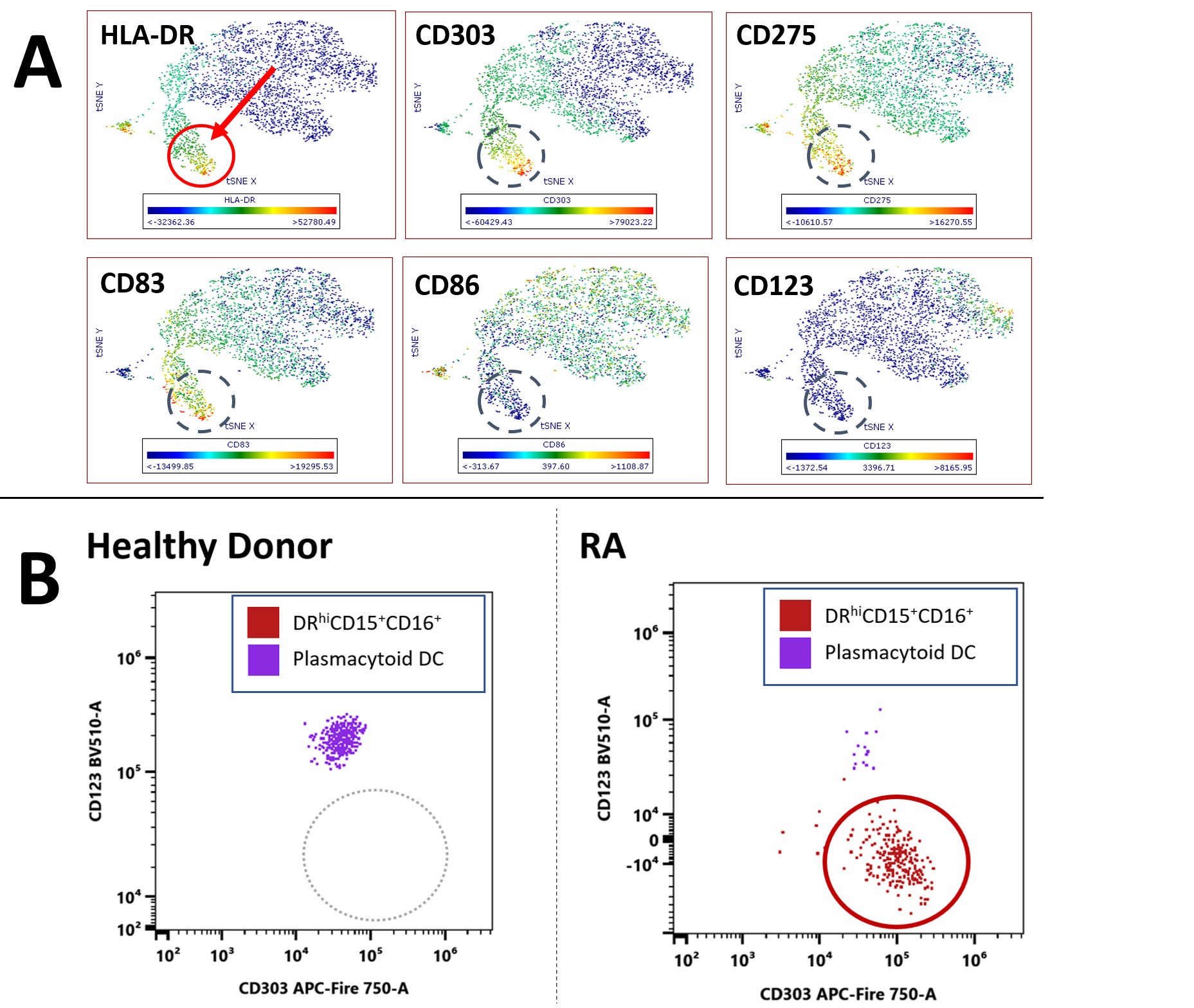
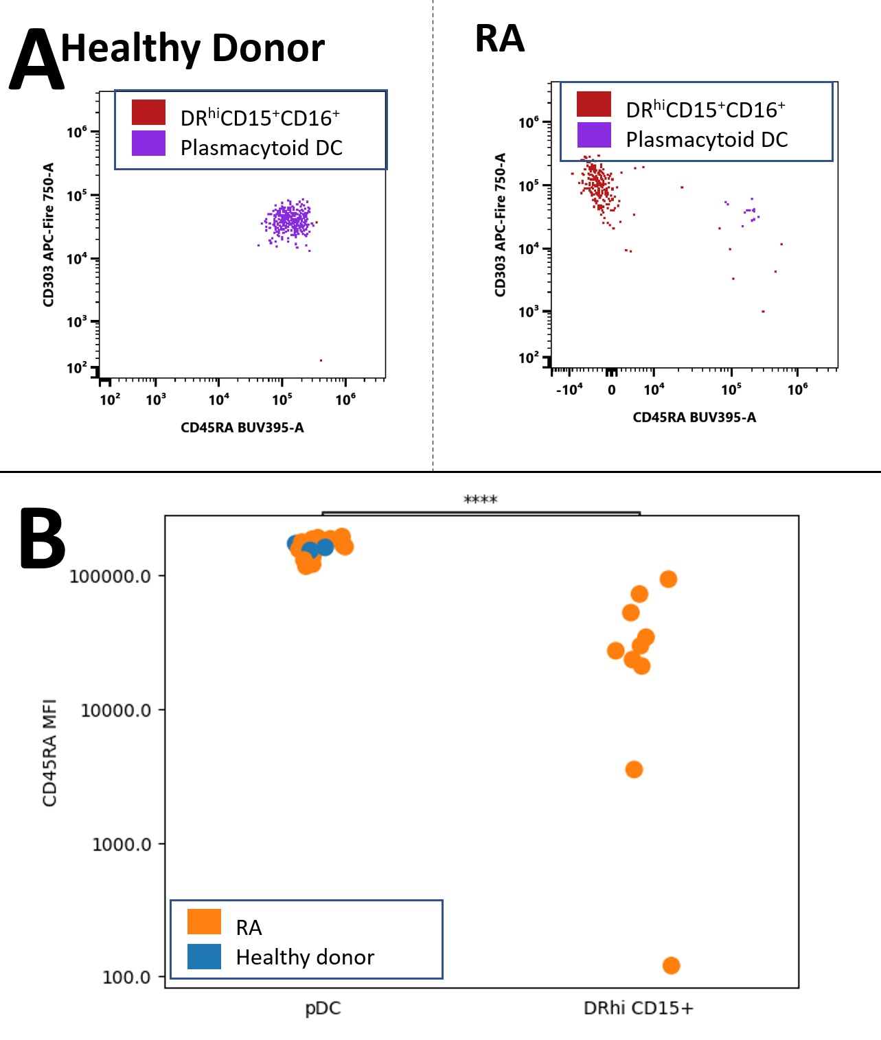
Results: In RA we found a non-lymphoid DRhi CD15+ population that was virtually absent in healthy donors (HD)(RA 0.98% vs. HD 0.05% of non-lymphoid DRhi; p<0.01, Fig. 1B, 2B); these cells have near uniform CD16 co-expression (Fig 1A, gated in last plot).DRhi CD15+ showed—as expected from granulocytic cells — high side scatter but differed from granulocytes by a striking co-expression of plasma cytoid (pDC) marker CD303 (Fig. 2A, B; circled);CD123, highly expressed by pDC, was not expressed. Given the shared features with both granulocytes and pDC we refer to this population as DRhi 'Hybrid' cells. DRhi Hybrids formed a separate cluster in RA which, along with CD303, co-expressed CD83 and CD275 (ICOS-L) (Fig. 2A, circled population).Lack of CD45RAseparatedCD303+DRhi Hybrids from their apparent bone fide CD303+ CD45RAint pDC counterparts (Fig. 3A and B, p< 0.001), CD45RA expression of RA pDC and HD pDC did not differ (Fig 3B, left). CCR3 expression within non-lymphoid DRhi was negligible (not shown).
Conclusion: RA blood contains DRhiCD15+ cells, contributing to the non-lymphoid DRhi pool in RA. Because these low-density cells share features with both granulocytes and pDCs we refer to them as DRhi Hybrids; their expression of CD303 challenges the notion that this molecule is specific for plasmacytoid DC. Co-expression of CD83, CD275 suggests an inflammatory potential whereas their lack of CD45RA (as opposed to CD303+pDCs) is reminiscent of the altered CD45RA expression we found previously in a DC2-like DRhi subset. Joint expression of CD303 and DR suggests that DRhi Hybrids maybe functionally involved in the capture and ultimate presentation of RA self-peptides to T lymphocytes. These apparently new DRhi Hybrids and other unorthodox DRhi populations are potential treatment targets in RA.
Flow cytometry and quantification of non-lymphoid DRhiCD15+ in RA. A) Gating strategy, patient with severe RA shown. B) DRhiCD15+ quantification in healthy donors (HD; blue) and RA (orange) and as percentage of non-lymphoid DRhi, logarithmic scale. Kruskal-Wallis Test ** p=0.0063
Non-lymphoid DRhiCD15+CD16+. A) t-SNE from an RA patient with debilitating polyarthritis population (red circle and arrow) highlighting co-expression of CD303, CD83, CD275; lack of CD123. Red: high expression. Blue: low expression. B) Bi-axial gating of DRhiCD15+CD16+ and pDC reference populations. CD303 x-axis, CD123 y-axis. Left plot: healthy donor Right plot: RA patient.
Comparison of CD303+ DRhiHybrids with CD303+ plasmacytoid DC. A) CD45RA and CD303 expression in healthy donor plasmacytoid DC (left; purple) and RA (with presence of DRhi hybrids) B) Quantification of CD45RA MFI of CD303+ pDC from RA (orange) and HD (blue) and CD303+ DRhi Hybrids. logarithmic scale. Kruskal-Wallis Test **** p=0.0008324
C. Geier: None; H. Qudsi: None; J. BenGabr: None; R. Winchester: None; A. Perl: None.