Session: (1734–1775) RA – Etiology and Pathogenesis Poster
1760: Methotrexate Augments the Release of Granulocyte-macrophage Colony-stimulating Factor from Activated Rheumatoid Arthritis Fibroblast-like Synoviocytes - Possible Consequences for Persistence of Joint Inflammation
Beatrice Bergström1, Tilia Selldén1, Miriam Bollmann2, Mattias N.D Svensson1 and Anna-Karin Hultgård Ekwall3, 1University of Gothenburg, Gothenburg, Sweden, 2Department of Rheumatology and Inflammation research, Sahlgrenska Academy, University of Gothenburg, Gothenburg, Sweden, 3University of Gothenburg, Kullavik, Sweden
Background/Purpose: Activated fibroblast-like synoviocytes (FLS) are important mediators of synovitis and structural damage in rheumatoid arthritis (RA)[1]. Granulocyte-macrophage colony-stimulating factor (GM-CSF, encoded by the CSF2 gene) is released from activated RA-FLS and promotes the production of pro-inflammatory cytokines from macrophages, which in turn activates FLS creating a viscous cycle[2]. We investigated the effects of anti-rheumatic drugs on gene expression, in particular RA risk genes such as CSF2, in activated RA-FLS, and on cytokine release from RA synoviocyte cultures.
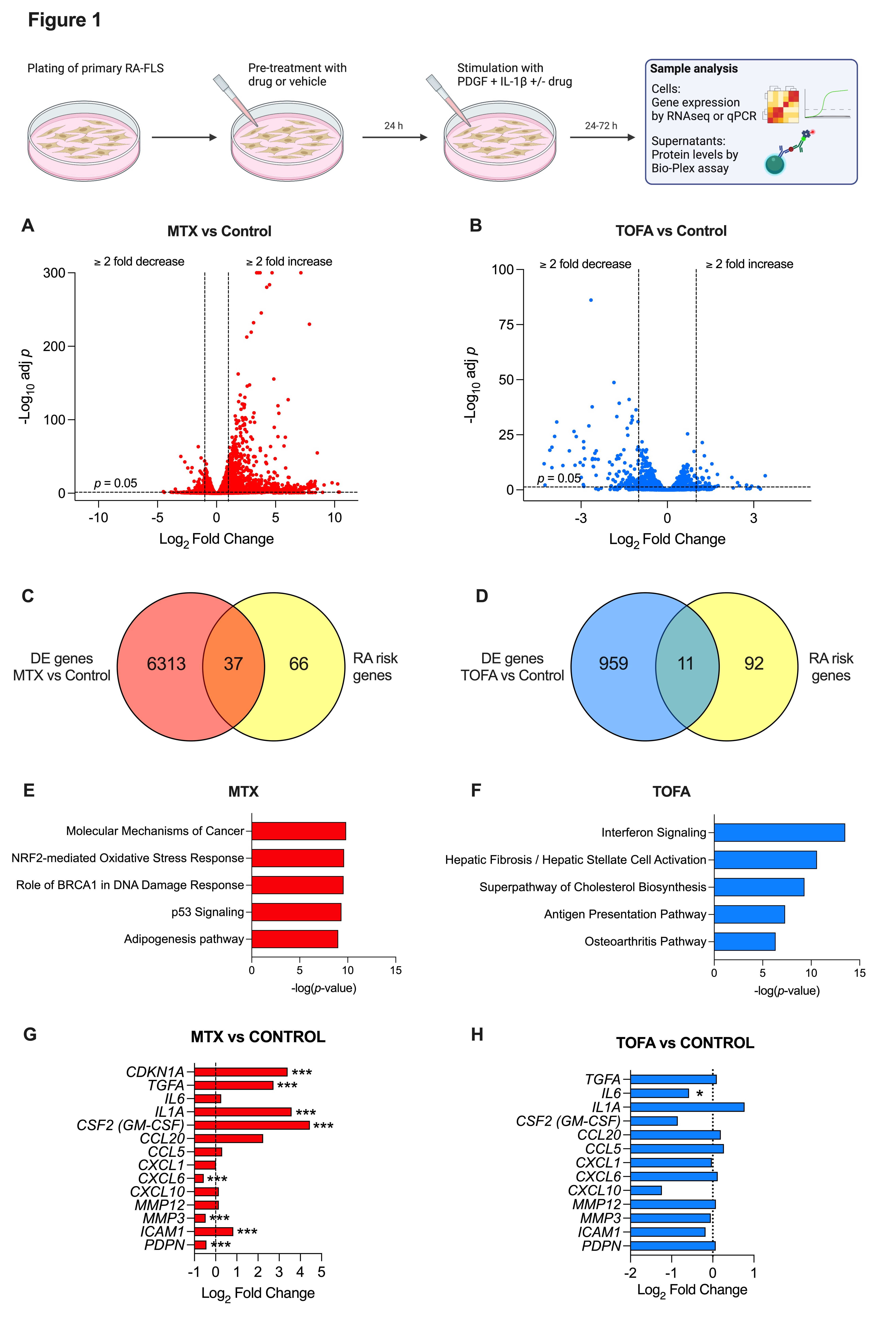
Methods: Primary FLS were established from synovial tissue collected from patients with RA undergoing arthroplasty or synovial biopsy. FLS were pre-treated with methotrexate (MTX), tofacitinib (TOFA, janus kinase inhibitor), zimlovisertib (IRAK4 inhibitor) or vehicle followed by stimulation with platelet-derived growth factor (PDGF) and interleukin-1β (IL-1β) for 24-72 hours (Fig.1, top). An RA ex-vivo synovial bioassay was performed according to Kuo et al.[3]. Cells were collected for RNAseq or qPCR, and supernatants were analyzed for protein concentrations using Bio-Plex assay.
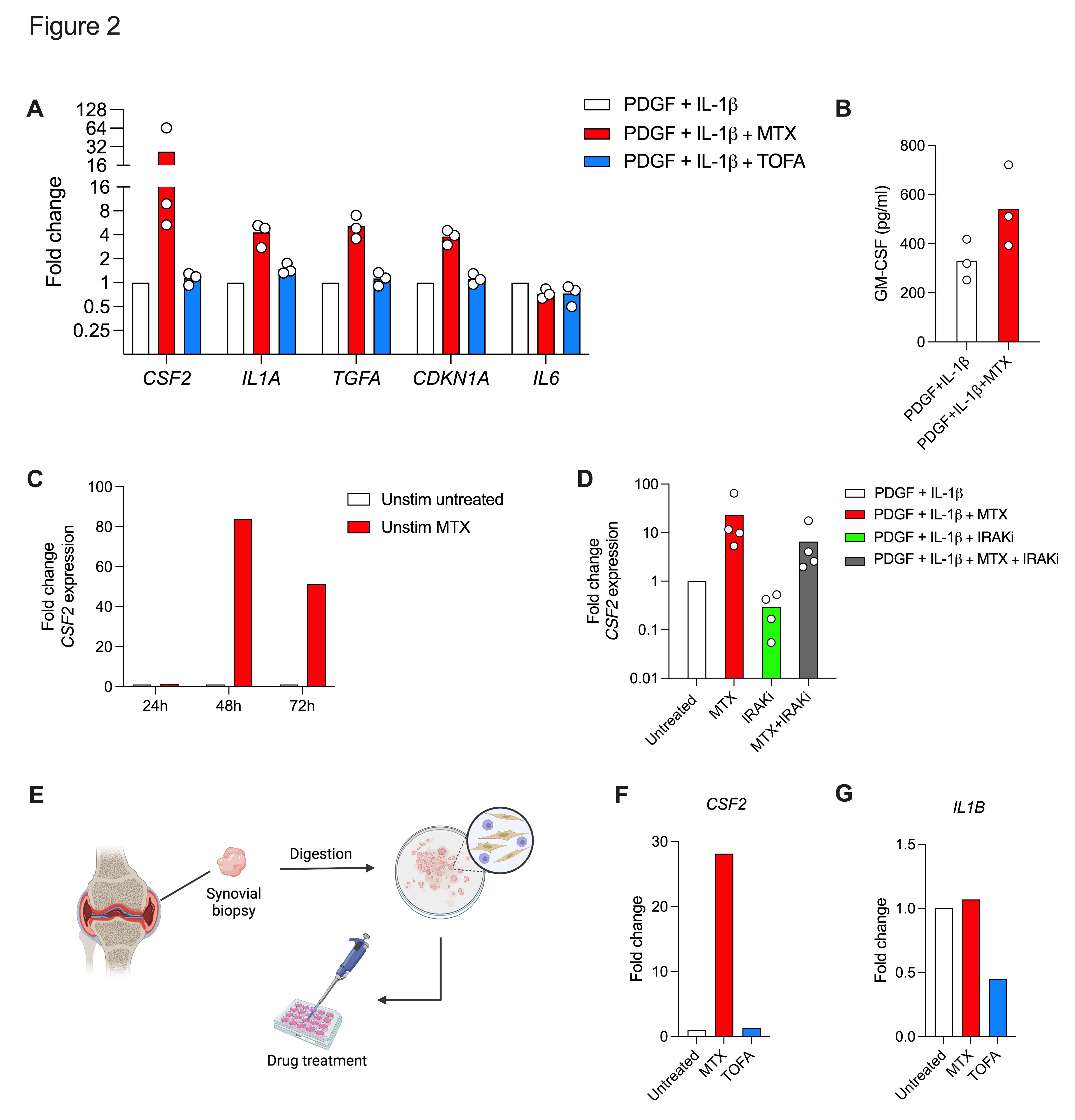
Results: MTX demonstrated an overall activation of gene expression in PDGF+IL-1β stimulated RA-FLS compared to untreated (6350 differentially expressed (DE) genes), and to TOFA treated (970 DE genes) samples (Fig. 1A-B). MTX had a greater effect on RA risk genes (35 % DE in MTX vs CON) compared to TOFA (10 % DE in TOFA vs CON) (Fig.1C-D). Pathway analysis showed largest effects on Molecular mechanisms of cancer (MTX) and Interferon signaling (TOFA) respectively (Fig. 1E-F). Targeted analysis revealed that MTX in addition to the known induction of CDKN1A (p21)[4], profoundly increased CSF2, IL1A and TGFA expression compared to control treated activated FLS (Fig.1G). TOFA reduced IL6 and CXCL10 expression as expected (Fig.1H). RNAseq data were confirmed by qPCR and on protein level in FLS supernatants (Fig.2A-B). Furthermore, MTX induced CSF2 expression also in unstimulated RA-FLS (Fig.2C). IRAK4i could not prevent the increased GM-CSF expression by MTX in stimulated RA-FLS (Fig.2D). MTX induced CSF2 expression in RA ex-vivo synovial cultures (Fig. 2E-F) and failed to reduce IL-1β expression as opposed to TOFA (Fig.2G).
Conclusion: Methotrexate treatment augments the release of GM-CSF from RA-FLS and leads to sustained cytokine production from synoviocytes. This off-target effect might contribute to the persistence of synovitis.
Primary RA-FLS were pre-treated with MTX, TOFA, or vehicle at 1-2,5 uM for 24 hours followed by stimulation with 20 ng/mL PDGF-BB and 2 ng/mL IL-1beta in medium with the drug or vehicle for 48 hours and finally processed for RNAseq. Vulcano plots show significantly DE genes by DESeq2 (p≤0.05) for MTX (A) and TOFA (B) vs untreated control. Venn diagram demonstrate effects of MTX (C) and TOFA (D) on RA risk genes identified by Genome wide association studies. Ingenuity Pathway Analysis revealed the five most significant pathways for MTX (E) and TOFA (F). Effects of MTX and TOFA on a core set of RA-associated FLS-expressed genes are demonstrated in G and H respectively. *Adjusted p ≤ 0.05, **p ≤ 0.01, ***p ≤ 0.001 (Benjamini-Hochberg).
Primary RA-FLS were pre-treated, stimulated and treated as outlined according to experimental design in figure 1. Graphs show fold change to untreated control by qPCR. A) Expression of RA-associated genes in stimulated RA-FLS treated with MTX (red bars) and TOFA (blue bars). B) GM-CSF concentration by Bio-Plex assay in supernatants of cells in A). C) Time course of CSF2 expression in MTX treated unstimulated RA-FLS. D) CSF2 expression in MTX, zimlovisertib (IRAK4i) or combination treated stimulated RA-FLS. E) Work flow of RA ex-vivo synovial bioassay. F) Induction of CSF2 expression by MTX and G) expression of IL-1beta in synoviocyte cultures (Bio-Assay) treated with MTX (red) and TOFA (blue).
B. Bergström: None; T. Selldén: None; M. Bollmann: None; M. Svensson: None; A. Hultgård Ekwall: AbbVie/Abbott, 1, 2, Boehringer-Ingelheim, 6, Pfizer, 1.