Camilla R. L. Machado1, David Boyle1, Narayanan B. Perumal2, Robert J. Benschop3 and Gary Firestein1, 1University of California San Diego, San Diego, CA, 2Eli Lilly and Company, San Diego, CA, 3Eli Lilly and Company, Indianapolis, IN
Background/Purpose: The synovial lining of rheumatoid arthritis (RA) is formed by a network of fibroblast-like synoviocytes (FLS). Cadherins are type I transmembrane proteins and participate in FLS cell-to-cell contacts, particularly cadherin 11. Unbiased profiling of cadherin expression in FLS using epigenetic data identified cadherin 6 (CDH6) as a gene of interest in RA FLS. CDH6, which is a type II cadherin, has mainly been studied in oncology where high expression is associated with cancer progression and invasion. CDH6 in FLS could regulate similar functions in RA. To understand the diverse functions of CDH6 in FLS, we analyzed its cellular distribution in synovial tissue.
Methods: RA and osteoarthritis (OA) synovial tissue (n=6 each) were obtained at arthroplasty. RA FLS and OA FLS lines (n=6 each) were derived from enzymatically dispersed synovial tissue and used from passages 5-7. Western blot analysis was used for protein quantification with anti-CDH6 antibody and was normalized to GAPDH. The distribution of CDH6 in FLS and synovial tissue was evaluated by immunofluorescence and confocal microscopy.
Results: Initial immunofluorescence studies of RA synovial tissue revealed abundant CDH6 in the intimal lining. Using anti-CD68 and anti-CDH6 antibodies in double stained tissues, we observed CDH6 expression in lining macrophages and fibroblasts. CDH6 was also expressed to a lesser degree in the sublining, particularly in the perivascular regions and included macrophages and non-macrophages (Figure 1). A similar distribution of CDH6 protein was observed in OA synovium and suggests that CDH6 is an important component in the synovial architecture. Because FLS reside in the intimal lining and were strongly CDH6 positive, we explored the expression and distribution of CDH6 in cultured FLS. Western blots showed that expression is higher in RA than OA FLS (1.2±0.01 vs 0.1±0.01 OD, p=0.03; n=4 each). Using immunostaining, expression was higher in FLS cultured at higher cell density and was ~2-fold higher in confluent vs. non-confluent cultures. Thus, CDH6 might be regulated, in part, by cell-cell contact. More intriguing, confocal microscopy showed significant expression of CDH6 protein in the cytoplasm, peri-nuclear regions, and nucleus in addition to the cell membrane.
Conclusion: CDH6 is expressed in synovial lining and sublining fibroblasts and macrophages. The striking intracellular distribution suggests additional functions beyond adhesion and homotypic aggregation, such as signalling and regulation of gene transcription. Thus, CDH6 has similarities to E-Cadherin, which also localizes to intracellular structures and participates in biologic functions. Our data suggest for the first time that CDH6 might have multiple functions in FLS that contribute to its pathogenic behaviour.
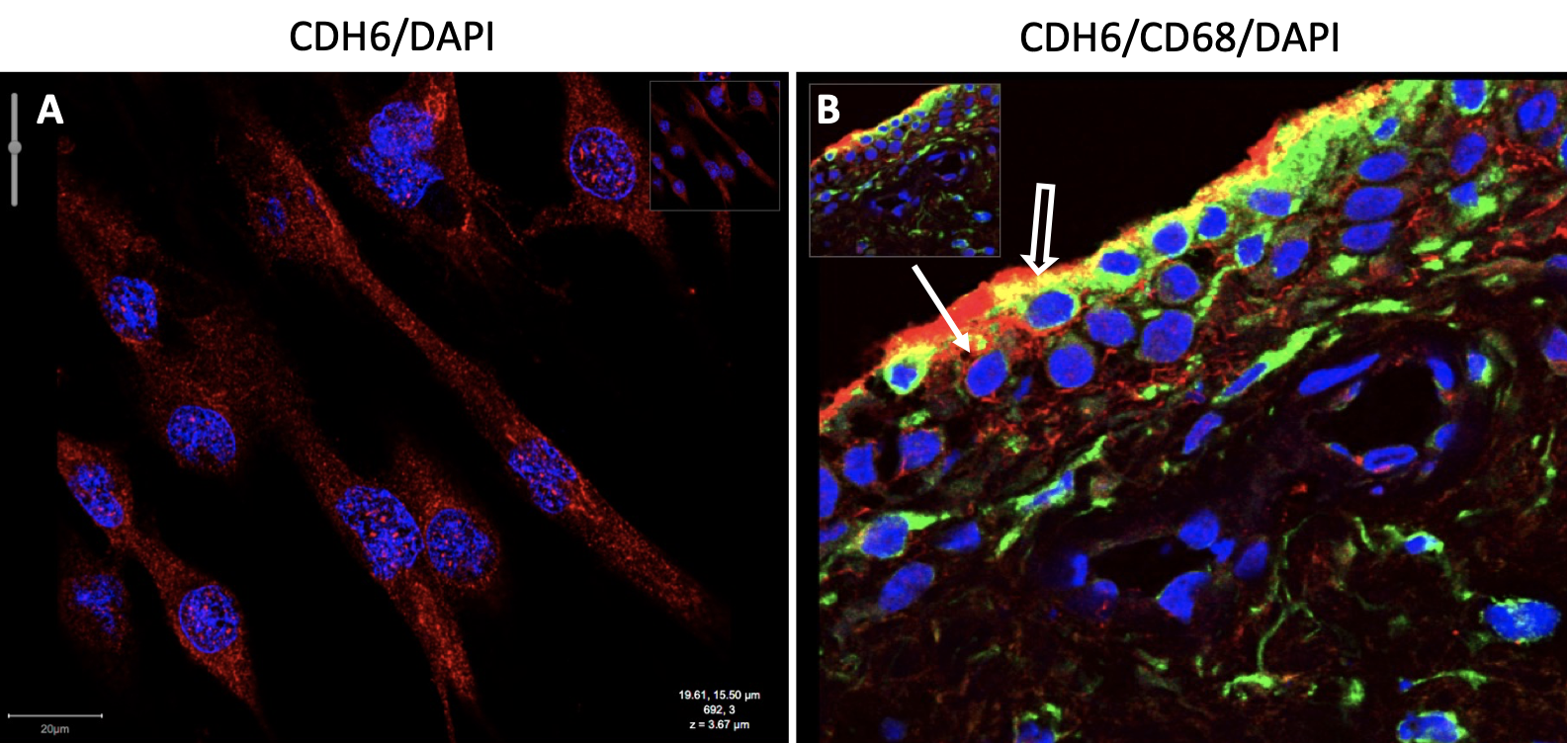
Figure 1. CDH6 expression in RA FLS and RA synovium. We performed confocal microscopy to determine if CDH6 is present in the synovial lining and its distribution in RA FLS. A) Representative image of RA FLS with anti-CDH6 (red) and DAPI (blue). CDH6 is found in the cytoplasm, peri-nuclear regions, nucleus, and the cell membrane. B) Representative image of RA synovium with anti-CDH6 (red) and -CD68 (green) (macrophage) antibodies show lining and sublining expression of CDH6, most likely in FLS (solid white arrow) and lining macrophages (open white arrow).
C. R. L. Machado: Eli Lilly, 5; D. Boyle: None; N. B. Perumal: Eli Lilly, 3; R. J. Benschop: Eli Lilly and Company, 3, 11; G. Firestein: Eli Lilly, 5.