Poster Session C
Rheumatoid arthritis (RA)
Session: (1734–1775) RA – Etiology and Pathogenesis Poster
1749: Fatty Acid Synthase Is a Critical Repressor of Ferroptosis in Rheumatoid Arthritis
Tuesday, November 14, 2023
9:00 AM - 11:00 AM PT
Location: Poster Hall

Qi Cheng, DO (he/him/his)
The Second Affiliated Hospital of Zhejiang University School of Medicine
Hangzhou, Zhejiang, ChinaDisclosure information not submitted.
Abstract Poster Presenter(s)
Qi Cheng, Mo chen, Xin chen, Yifan Xie, Yan Du and Huaxiang wu, The Second Affiliated Hospital Of Zhejiang University School Of Medicine, Hangzhou, China
Background/Purpose: Rheumatoid arthritis (RA) is a systemic autoimmune disease with synovial inflammation as the main pathological feature, and can eventually lead to irreversible joint or organ damage. Recently, several studies have reported that ferroptosis was involved in the pathogenesis of RA. However, the role of ferroptosis in abnormal activation of RA synovial fibroblasts (RASFs) is poorly understood. The purpose of our study was to explore the effect of fatty acid synthase (FASN) on ferroptosis in RASFs.
Methods: FASN expression was assessed by quantitative polymerase chain reaction (qPCR), western blotting and immunohistochemistry in synovial tissues and SFs from RA patients, osteoarthritis (OA) patients and collagen induced arthritis (CIA) mice. SF activation was evaluated by qPCR, CCK-8 and wound healing assay. Lipid peroxidation was detected by MDA levels and C11-BODIPY 581/591 fluorescence intensity. and transient small interfering RNA knockdown were performed to examine expression of SLC7A11 and signaling pathway protein.
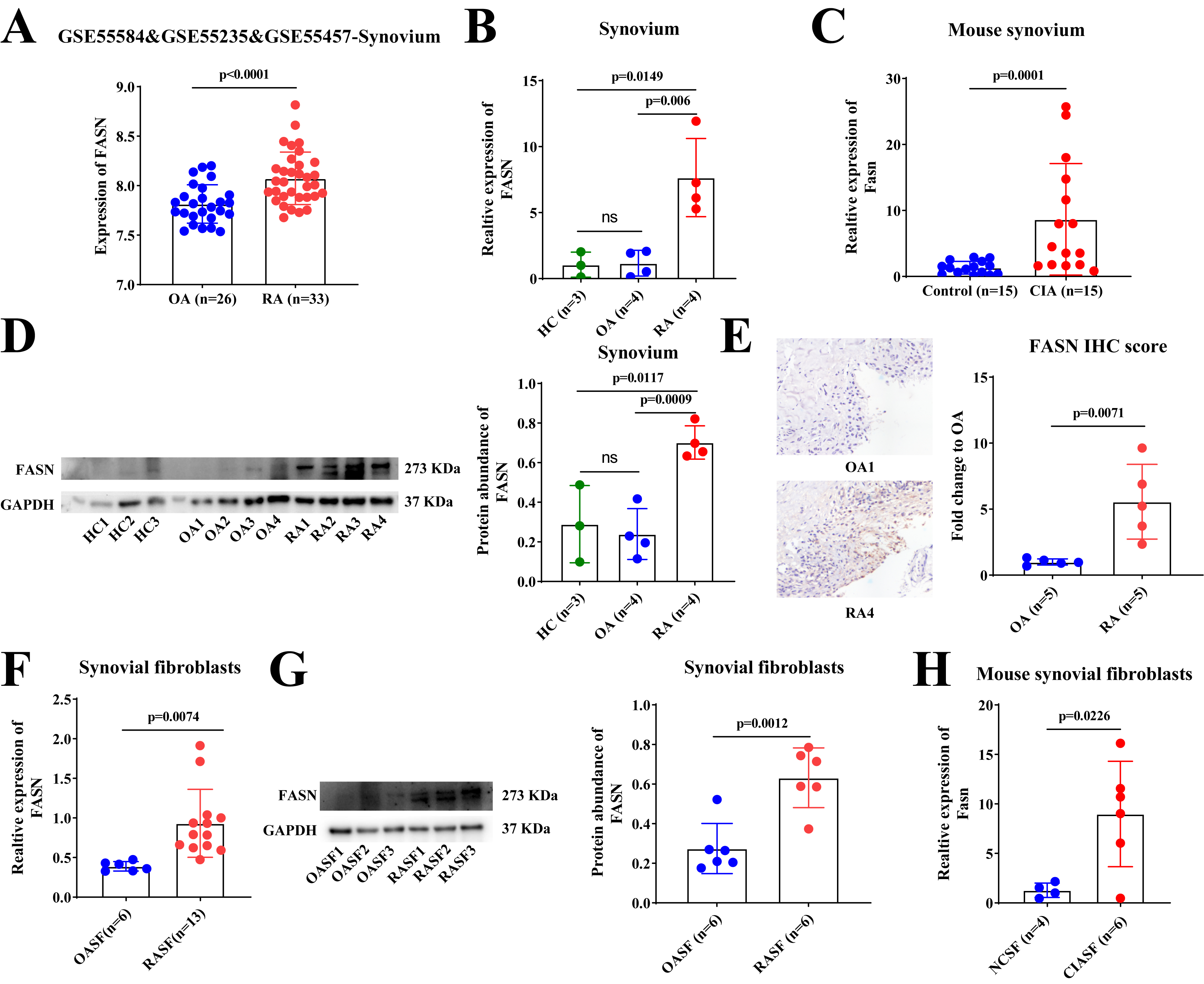
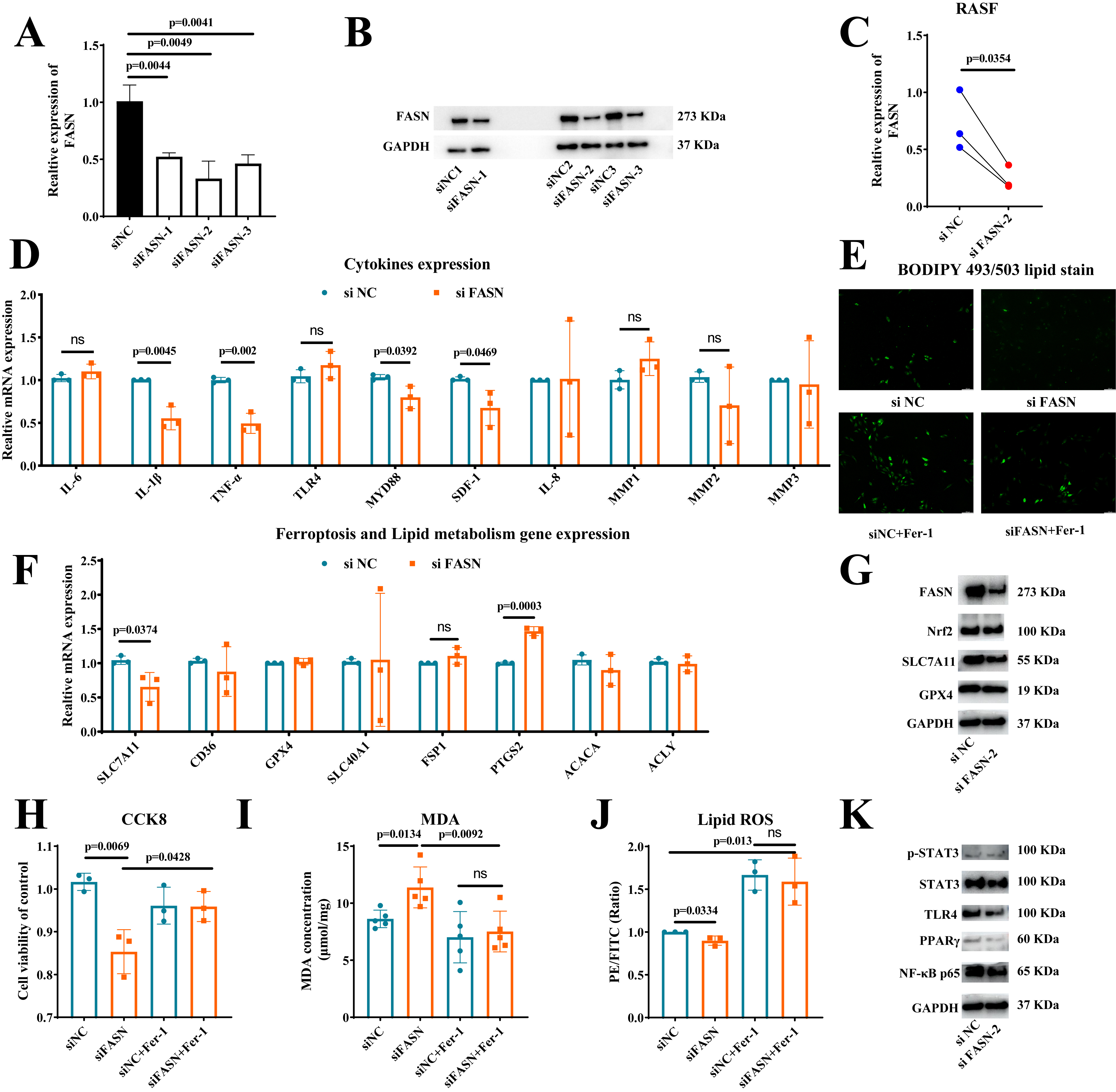
Results: FASN was found to be significantly higher expressed in synovial tissue and SF from RA patients and CIA mice (Figure 1). Silencing of FASN by siRNA in RASFs reduced expression of FASN, SLC7A11, inflammatory cytokines (IL-1β, TNF-α and SDF-1), level of lipid and cell viability, and enhanced the expression of PTGS2 and level of lipid peroxidation, but not affected the expression of other ferroptosis (SLC40A1, FSP1 and GPX4) and lipid (ACACA, ACLY and CD36) genes (Figure 2A-2J). Moreover, treatment with the ferroptosis inhibitor, Ferrostatin-1 (Fer-1) could reverse the effect of FASN knockdown on lipid synthesis, cell viability and ferroptosis in RASFs (Figure 2E-2J). Mechanistically, FASN may promote inflammatory cytokines expression by TLR4/MYD88/NF-κ B signaling pathway, and enhance SLC7A11 expression by phosphorylating the STAT3 signal to suppress ferroptosis of RASFs (Figure 2K).
Conclusion: Our study found that FASN was a ferroptosis suppressor in RASFs. Silencing of FASN inhibited the abnormal activation of RASFs by suppressing STAT3/SLC7A11 axis and the TLR4/MYD88/NF-κB signaling pathway. Target FASN and ferroptosis may serve as a promising novel therapeutic strategy for RA.


Q. Cheng: None; M. chen: None; X. chen: None; Y. Xie: None; Y. Du: None; H. wu: None.
Background/Purpose: Rheumatoid arthritis (RA) is a systemic autoimmune disease with synovial inflammation as the main pathological feature, and can eventually lead to irreversible joint or organ damage. Recently, several studies have reported that ferroptosis was involved in the pathogenesis of RA. However, the role of ferroptosis in abnormal activation of RA synovial fibroblasts (RASFs) is poorly understood. The purpose of our study was to explore the effect of fatty acid synthase (FASN) on ferroptosis in RASFs.
Methods: FASN expression was assessed by quantitative polymerase chain reaction (qPCR), western blotting and immunohistochemistry in synovial tissues and SFs from RA patients, osteoarthritis (OA) patients and collagen induced arthritis (CIA) mice. SF activation was evaluated by qPCR, CCK-8 and wound healing assay. Lipid peroxidation was detected by MDA levels and C11-BODIPY 581/591 fluorescence intensity. and transient small interfering RNA knockdown were performed to examine expression of SLC7A11 and signaling pathway protein.
Results: FASN was found to be significantly higher expressed in synovial tissue and SF from RA patients and CIA mice (Figure 1). Silencing of FASN by siRNA in RASFs reduced expression of FASN, SLC7A11, inflammatory cytokines (IL-1β, TNF-α and SDF-1), level of lipid and cell viability, and enhanced the expression of PTGS2 and level of lipid peroxidation, but not affected the expression of other ferroptosis (SLC40A1, FSP1 and GPX4) and lipid (ACACA, ACLY and CD36) genes (Figure 2A-2J). Moreover, treatment with the ferroptosis inhibitor, Ferrostatin-1 (Fer-1) could reverse the effect of FASN knockdown on lipid synthesis, cell viability and ferroptosis in RASFs (Figure 2E-2J). Mechanistically, FASN may promote inflammatory cytokines expression by TLR4/MYD88/NF-κ B signaling pathway, and enhance SLC7A11 expression by phosphorylating the STAT3 signal to suppress ferroptosis of RASFs (Figure 2K).
Conclusion: Our study found that FASN was a ferroptosis suppressor in RASFs. Silencing of FASN inhibited the abnormal activation of RASFs by suppressing STAT3/SLC7A11 axis and the TLR4/MYD88/NF-κB signaling pathway. Target FASN and ferroptosis may serve as a promising novel therapeutic strategy for RA.

Figure 1 Expression of FASN in synovial tissues and synovial fibroblasts extracted from RA patients and CIA model mice. A. Expression of FASN in 33 RA and 26 OA synovial tissues from GSE55584&GSE55325&GSE55457 dataset. B-C. Expression of FASN in synovial tissues extracted from RA patients (B) and CIA model mice (C). D. FASN was detected by western blot in synovial tissues from 3 HC, 4 OA and 4 RA patients. E. FASN was detected by immunohistochemistry in synovial tissues from 5 OA and 5 RA patients. F-G, FASN was detected by PCR (F) and western blot (G) in SFs. H. Fasn was detected by PCR in mouse SFs. Values of P < 0.05 were considered significant. SFs, synovial fibroblasts. RA, rheumatoid arthritis; OA, osteoarthritis; SFs, synovial fibroblasts.

Figure 2 The role of FASN in production of inflammatory cytokines, lipid and regulation of ferroptosis on RASFs. A-B. Silencing efficiency of siRNA targeting FASN (siFASN) was detected by qPCR (A) and Western blot (B). C. mRNA level of FASN in 3 SFs after knockdown by siRNA. D. Expression of cytokines in SFs after transfection of siRNA to knockdown FASN. E. Representative fluorescent images of BIOIPY 493/503 lipid statin. F. Expression of ferroptosis and lipid genes in SFs after transfection of siRNA to knockdown FASN. G. Protein abundance of FASN, SLC7A11, Nrf2 and GPX4 in SFs after transfection of siRNA to knockdown FASN. H. Cell viability of SFs detected by a Cell Counting Kit-8. I. Intracellular MDA levels in SFs detected by a Lipid Oxidation (MDA) Assay Kit SFs. J. Fluorescence intensity of C11-BODIPY 581/591 in SFs detected by flow cytometry. K. Protein abundance of p-STAT3, STAT3, TLR4, PPARγ and NF-κB p65 in SFs after transfection of siRNA to knockdown FASN. Values of P < 0.05 were considered significant.
Q. Cheng: None; M. chen: None; X. chen: None; Y. Xie: None; Y. Du: None; H. wu: None.


