Poster Session B
Vasculitis
Session: (1554–1578) Vasculitis – Non-ANCA-Associated & Related Disorders Poster II
1558: Development of Posterior Uveitis in Behçet’s Syndrome Patients with Vitreous Cells Without Any Other Posterior Involvement
Monday, November 13, 2023
9:00 AM - 11:00 AM PT
Location: Poster Hall
- SE
Sinem Esatoglu, MD
Istanbul University-Cerrahpasa, Cerrahpasa Medical Faculty, Department of Internal Medicine, Division of Rheumatology
İstanbul, TurkeyDisclosure information not submitted.
Abstract Poster Presenter(s)
Didar Ucar1, Basak Ecem Bircan2, Nigar Rustamli2, Bilge Batu Oto1, Vedat Hamuryudan3, Sinem Nihal Esatoglu3 and Gulen Hatemi3, 1Istanbul University-Cerrahpasa, Cerrahpasa Medical School, Department of Ophthalmology, Istanbul, Turkey, 2Istanbul University-Cerrahpasa, Cerrahpasa Medical School, Department of Internal Medicine, Istanbul, Turkey, 3Istanbul University-Cerrahpasa, Cerrahpasa Medical School, Department of Internal Medicine, Division of Rheumatology, Istanbul, Turkey
Background/Purpose: A considerable number of patients with Behçet's syndrome (BS) have vitreous cells (VC) on slit lamp examination at the time of diagnosis. However, the prognostic importance of VC and their association with the development of posterior uveitis (PU) requiring immunosuppressive treatment is unknown. We aimed to determine the prognostic importance of VC in BS patients.
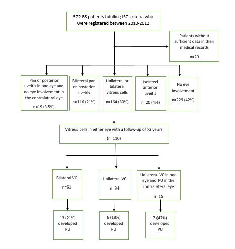
Methods: The charts of 572 consecutive BS patients fulfilling ISG criteria who were registered between 2010 and 2012 were reviewed. At baseline visit 164 patients had VC in one or both eyes. Among the remaining patients, 229 had no eye involvement, 116 patients had bilateral pan or posterior uveitis, 14 had unilateral pan or posterior uveitis and no eye involvement in the other eye, 20 had isolated anterior uveitis, and 29 had insufficient data in their medical records. Among the 164 patients with VC, 110 patients with a follow-up of ≥2 years were included in this study (Figure).
Results: At baseline, among the 110 included patients (68 men, mean ± SD age: 31±9 years), 61 had VC in both eyes, 34 had VC in only one eye, and 15 had VC in one eye and PU in the other eye. There was anterior uveitis (AU) in addition to VC in the same eye in 13 patients at baseline.
New PU developed in 26 (24%) patients during a mean follow-up of 1.9±1.1 years. Seven patients that developed PU in the eye with VC had had PU in the contralateral eye at baseline. This means 7 of 15 patients with VC in one eye and PU in the contralateral eye developed bilateral PU despite treatment. Two of these 7 patients had developed AU before they developed PU. Additionally, 6 patients that developed PU in the eye with VC had anterior uveitis in the same eye at baseline (Figure).
Multivariate logistic regression analysis showed that the presence of both VH and AU in the same eye at baseline (OR, 3.75, 95% CI; 1.07-13.18) and the presence of VH in one eye and PU in the other eye at baseline (OR, 3.86, 95% CI; 1.19-12.60) were the independent risk factors for the development of PU in patients with VH. Age at diagnosis, sex, and the use of immunosuppressive medications during the first 3 years of follow-up were not associated with the development of PU.
Conclusion: Careful follow-up is required for patients with VC since one quarter developed PU within 2 years. The presence of PU in the contralateral eye and AU in the same eye are risk factors for the development of PU in patients with VC.

D. Ucar: None; B. Bircan: None; N. Rustamli: None; B. Batu Oto: None; V. Hamuryudan: None; S. Esatoglu: None; G. Hatemi: None.
Background/Purpose: A considerable number of patients with Behçet's syndrome (BS) have vitreous cells (VC) on slit lamp examination at the time of diagnosis. However, the prognostic importance of VC and their association with the development of posterior uveitis (PU) requiring immunosuppressive treatment is unknown. We aimed to determine the prognostic importance of VC in BS patients.
Methods: The charts of 572 consecutive BS patients fulfilling ISG criteria who were registered between 2010 and 2012 were reviewed. At baseline visit 164 patients had VC in one or both eyes. Among the remaining patients, 229 had no eye involvement, 116 patients had bilateral pan or posterior uveitis, 14 had unilateral pan or posterior uveitis and no eye involvement in the other eye, 20 had isolated anterior uveitis, and 29 had insufficient data in their medical records. Among the 164 patients with VC, 110 patients with a follow-up of ≥2 years were included in this study (Figure).
Results: At baseline, among the 110 included patients (68 men, mean ± SD age: 31±9 years), 61 had VC in both eyes, 34 had VC in only one eye, and 15 had VC in one eye and PU in the other eye. There was anterior uveitis (AU) in addition to VC in the same eye in 13 patients at baseline.
New PU developed in 26 (24%) patients during a mean follow-up of 1.9±1.1 years. Seven patients that developed PU in the eye with VC had had PU in the contralateral eye at baseline. This means 7 of 15 patients with VC in one eye and PU in the contralateral eye developed bilateral PU despite treatment. Two of these 7 patients had developed AU before they developed PU. Additionally, 6 patients that developed PU in the eye with VC had anterior uveitis in the same eye at baseline (Figure).
Multivariate logistic regression analysis showed that the presence of both VH and AU in the same eye at baseline (OR, 3.75, 95% CI; 1.07-13.18) and the presence of VH in one eye and PU in the other eye at baseline (OR, 3.86, 95% CI; 1.19-12.60) were the independent risk factors for the development of PU in patients with VH. Age at diagnosis, sex, and the use of immunosuppressive medications during the first 3 years of follow-up were not associated with the development of PU.
Conclusion: Careful follow-up is required for patients with VC since one quarter developed PU within 2 years. The presence of PU in the contralateral eye and AU in the same eye are risk factors for the development of PU in patients with VC.

Figure. Flowchart of the patients
D. Ucar: None; B. Bircan: None; N. Rustamli: None; B. Batu Oto: None; V. Hamuryudan: None; S. Esatoglu: None; G. Hatemi: None.


