Poster Session B
Myopathic rheumatic diseases (polymyositis, dermatomyositis, inclusion body myositis)
Session: (1155–1182) Muscle Biology, Myositis & Myopathies – Basic & Clinical Science Poster II
1164: Can We Differentiate Patients with Dysferlinopathies and Inflammatory Myopathies by Muscle Ultrasound? A Discriminant Analysis Study
Monday, November 13, 2023
9:00 AM - 11:00 AM PT
Location: Poster Hall
.png)
Carlos Pineda, PhD
Instituto Nacional de Rehabilitacion
Mexico City, Federal District, MexicoDisclosure information not submitted.
Abstract Poster Presenter(s)
Carina Soto-Fajardo1, Sinthia Solorzano Flores1, Abish Angeles-Acuña1, Fabian Carranza Enriquez1, Rosa-Elena Escobar-Cedillo2, Saul Renan-Leon2, Karina Arias Callejas3, Alejandra Enriquez-Luna2, Graciliano Ramon-Diaz2 and Carlos Pineda2, 1Instituto Nacional de Rehabilitación "Luis Guillermo Ibarra Ibarra", Mexico City, Mexico, 2Instituto Nacional de Rehabilitacion, Mexico City, Mexico, 3Instituto Nacional de Rehabilitación "Luis Guillermo Ibarra Ibarra", Mexico City, Mexico
Background/Purpose: Immune-mediated myopathies (IMM) are a heterogeneous group of diseases characterized by inflammation and muscle weakness; among their differential diagnoses are the dysferlinopathies, which are autosomal recessive neuromuscular disorders caused by mutations in the DYSF gene that present muscle weakness and significant increase of CK, just like IMM. The aim of this research is to determine the sonographic differences between dysferlinopathies and immune-mediated myopathies and whether these allow their classification.
Methods: Observational, cross-sectional, and analytical study in which we evaluated 20 muscles from 11 patients with dysferlinopathies and 11 with IMM. They were matched for age, sex, and time of disease evolution. Clinical and laboratory variables were analyzed. GE LOGIQTMe equipment with a 4-12 MHz linear transducer was used, and the thickness of each muscle was measured. A semiquantitative scale evaluated elementary lesions: atrophy, edema, power Doppler, and the Heckmatt scale (0-4) was calculated. Descriptive statistics were performed. Finally, discriminant analysis was performed to determine which ultrasound variables best predicted the diagnoses.
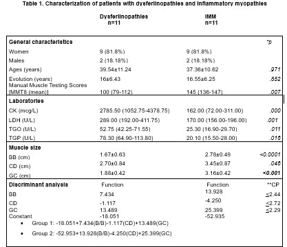
Results: A total of 40 muscles were evaluated, finding a greater degree of atrophy and a higher Heckmatt scale in patients with dysferlinopathies compared to MII (Table 1). Discriminant analysis showed that the set of 3 muscles, Right biceps/brachialis (BB), Right quadriceps (CD), and Gastrocnemius/right soleus (GC), had a diagnostic accuracy of 100% (sensitivity 100%, specificity 100%, canonical coefficient 0.733 p=.000). We present a set of 2 formulas that allow classifying with the highest score according to the measurement of the muscles in group 1 (dysferlinopathy) or group 2 (MII). Finally, a COR analysis was performed to determine the cut-off points of each muscle to classify as dysferlinopathies.
Conclusion: The study of 3 muscle groups (BB, CD, GC) presents high diagnostic accuracy in differentiating dysferlinopathies from MII, especially when no genetic study or antibodies are available and diagnostic doubt exists.

C. Soto-Fajardo: None; S. Solorzano Flores: None; A. Angeles-Acuña: None; F. Carranza Enriquez: None; R. Escobar-Cedillo: None; S. Renan-Leon: None; K. Arias Callejas: None; A. Enriquez-Luna: None; G. Ramon-Diaz: None; C. Pineda: None.
Background/Purpose: Immune-mediated myopathies (IMM) are a heterogeneous group of diseases characterized by inflammation and muscle weakness; among their differential diagnoses are the dysferlinopathies, which are autosomal recessive neuromuscular disorders caused by mutations in the DYSF gene that present muscle weakness and significant increase of CK, just like IMM. The aim of this research is to determine the sonographic differences between dysferlinopathies and immune-mediated myopathies and whether these allow their classification.
Methods: Observational, cross-sectional, and analytical study in which we evaluated 20 muscles from 11 patients with dysferlinopathies and 11 with IMM. They were matched for age, sex, and time of disease evolution. Clinical and laboratory variables were analyzed. GE LOGIQTMe equipment with a 4-12 MHz linear transducer was used, and the thickness of each muscle was measured. A semiquantitative scale evaluated elementary lesions: atrophy, edema, power Doppler, and the Heckmatt scale (0-4) was calculated. Descriptive statistics were performed. Finally, discriminant analysis was performed to determine which ultrasound variables best predicted the diagnoses.
Results: A total of 40 muscles were evaluated, finding a greater degree of atrophy and a higher Heckmatt scale in patients with dysferlinopathies compared to MII (Table 1). Discriminant analysis showed that the set of 3 muscles, Right biceps/brachialis (BB), Right quadriceps (CD), and Gastrocnemius/right soleus (GC), had a diagnostic accuracy of 100% (sensitivity 100%, specificity 100%, canonical coefficient 0.733 p=.000). We present a set of 2 formulas that allow classifying with the highest score according to the measurement of the muscles in group 1 (dysferlinopathy) or group 2 (MII). Finally, a COR analysis was performed to determine the cut-off points of each muscle to classify as dysferlinopathies.
Conclusion: The study of 3 muscle groups (BB, CD, GC) presents high diagnostic accuracy in differentiating dysferlinopathies from MII, especially when no genetic study or antibodies are available and diagnostic doubt exists.

C. Soto-Fajardo: None; S. Solorzano Flores: None; A. Angeles-Acuña: None; F. Carranza Enriquez: None; R. Escobar-Cedillo: None; S. Renan-Leon: None; K. Arias Callejas: None; A. Enriquez-Luna: None; G. Ramon-Diaz: None; C. Pineda: None.


