Poster Session A
Vasculitis
Session: (0691–0721) Vasculitis – Non-ANCA-Associated & Related Disorders Poster I
0707: Prevalence and Distribution of Vascular Calcifications at CT Scan in Patients with Large Vessel Vasculitis and Patients with Lymphoma: A Matched Cross-sectional Study
Sunday, November 12, 2023
9:00 AM - 11:00 AM PT
Location: Poster Hall
- CM
Chiara Marvisi, MD
University of Modena e Reggio Emilia
Reggio Emilia, Reggio Emilia, ItalyDisclosure information not submitted.
Abstract Poster Presenter(s)
Chiara Marvisi1, Giulia Besutti2, Pamela Mancuso3, Roberto Farì4, Filippo Monelli4, Matteo Revelli4, Rexhep Durmo5, Elena Galli6, Francesco Muratore7, Lucia Spaggiari2, Marta Ottone3, Stefano Luminari8, Pierpaolo Pattacini4, Paolo Giorgi Rossi3 and Carlo Salvarani9, 1IRCCS di Reggio Emilia, Reggio Emilia, Italy, Reggio Emilia, Italy, 2IRCCS REGGIO EMILIA, Reggio Emilia, Italy, 3Epidemiology Unit, Azienda Unità Sanitaria Locale-IRCSS Reggio Emilia, Reggio Emilia, Italy, 4Radiology Unit, Azienda Unità Sanitaria Locale-IRCCS Reggio Emilia, Reggio Emilia, Italy, 5Nuclear Medicine Unit, Azienda Unità Sanitaria Locale-IRCSS Reggio Emilia, Reggio Emilia, Italy, 6Azienda Unità Sanitaria Locale-IRCCS Di Reggio Emilia, Reggio Emilia, Italy, 7IRCCS di Reggio Emilia, Reggio Emilia, Italy, 8Ematology Unit, Azienda Unità Sanitaria Locale-IRCCS Reggio Emilia, Reggio Emilia, Italy, 9Azienda USL-IRCCS di Reggio Emilia, Reggio Emilia, Italy
Background/Purpose: Arterial wall calcifications are a hallmark of atherosclerosis and represent an important cardiovascular risk factor. Accelerated atherosclerosis and vascular calcifications have been reported in large vessel vasculitis (LVV), but data are scarce about the amount and localizations. The aim of this study was to compare the prevalence, entity, and local distribution of arterial wall calcifications evaluated on CT scans in patients with LVV and lymphoma.
Methods: All consecutive patients diagnosed with LVVs with available baseline PET-CT scan performed between 2007 and 2019 were included; lymphoma patients were matched by age (+/-5 years), sex, and year of baseline PET-CT (≤2013; >2013). CT images derived from baseline PET-CT scans of both patient groups were retrospectively reviewed by a single radiologist who, after setting a threshold of minimum 130 HU, semi-automatically computed vascular calcifications in three separate locations (coronaries, thoracic and abdominal arteries), quantified as Agatston and volume scores.
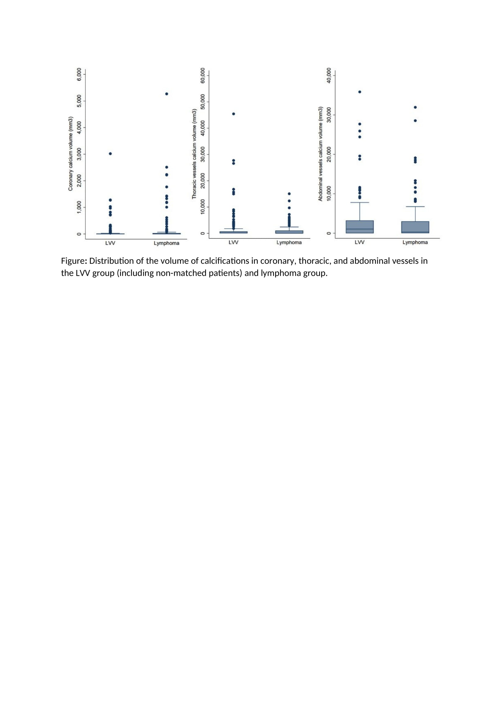
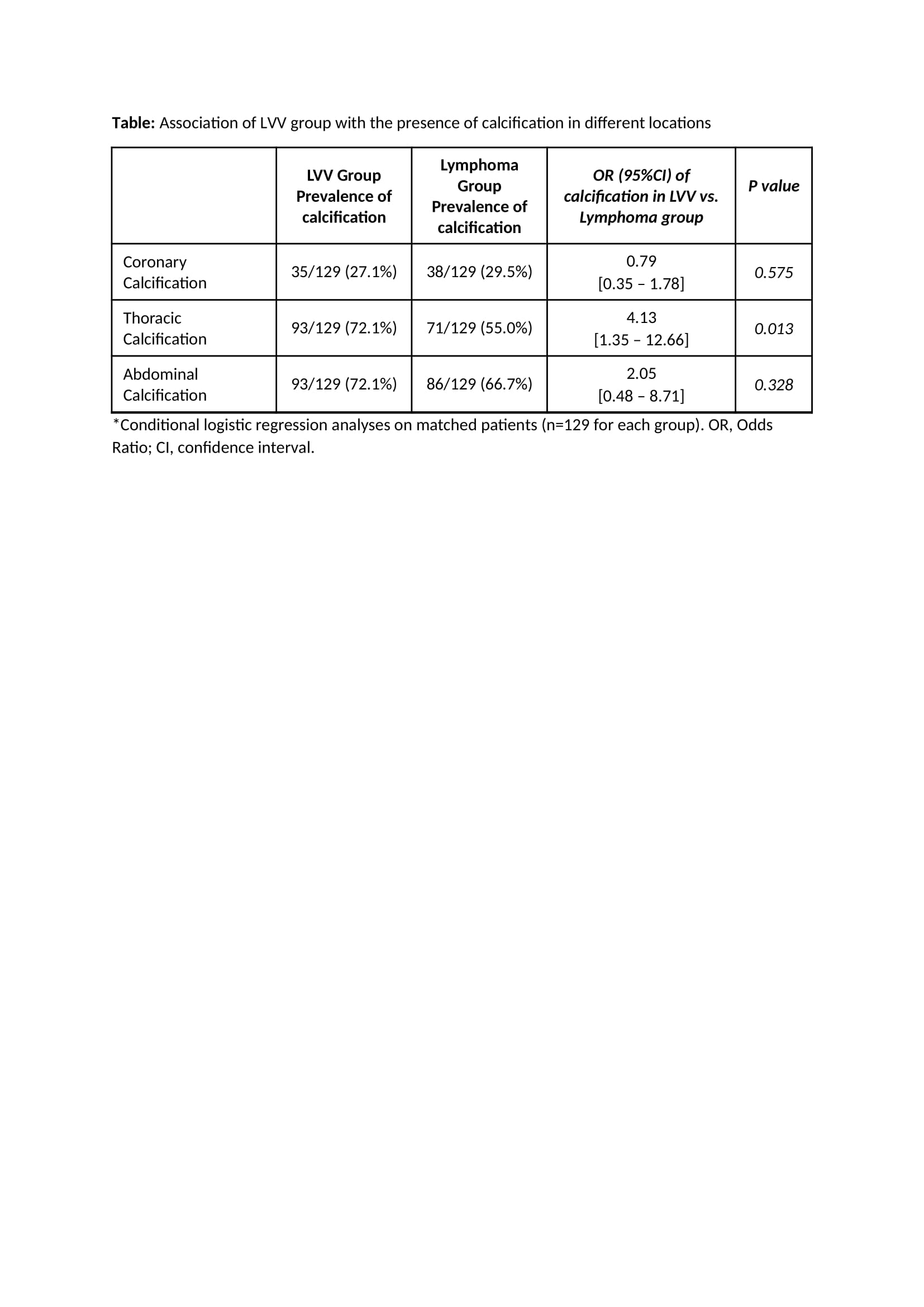
Results: A total of 266 patients were included (129 for each group and 8 non-matched LVV patients). Thoracic artery calcifications were more represented in LVV patients, when compared with lymphoma patients (mean volume 2026 in LVVs vs 1014 in lymphomas, p=0.054), coronary calcifications were higher in lymphoma patients (mean volume 104 in LVVs and 198 in lymphomas, p=0.13), whereas abdominal artery calcifications were equally distributed (mean volume 3220 in LVVs and 2712 in lymphomas) (Figure). Being in the LVVs group was associated with the presence of thoracic calcifications after adjusting by age and year of diagnosis (OR=4.13, 95%CI=1.35-12.66; p=0.013). Similarly, LVVs group was significantly associated with the volume score in the thoracic arteries (p=0.048) (Table).In patients >50 years old, calcifications in the coronaries were more extended in lymphoma patients (p=0.027 for volume).
Conclusion: When compared with lymphoma patients, LVVs patients have higher volumes of calcifications in the thoracic arteries, but not in coronary and abdominal arteries.


C. Marvisi: None; G. Besutti: None; P. Mancuso: None; R. Farì: None; F. Monelli: None; M. Revelli: None; R. Durmo: None; E. Galli: None; F. Muratore: None; L. Spaggiari: None; M. Ottone: None; S. Luminari: None; P. Pattacini: None; P. Giorgi Rossi: None; C. Salvarani: CSL Vifor, 1, 2, 6, Eli Lilly, 1, 2, 6.
Background/Purpose: Arterial wall calcifications are a hallmark of atherosclerosis and represent an important cardiovascular risk factor. Accelerated atherosclerosis and vascular calcifications have been reported in large vessel vasculitis (LVV), but data are scarce about the amount and localizations. The aim of this study was to compare the prevalence, entity, and local distribution of arterial wall calcifications evaluated on CT scans in patients with LVV and lymphoma.
Methods: All consecutive patients diagnosed with LVVs with available baseline PET-CT scan performed between 2007 and 2019 were included; lymphoma patients were matched by age (+/-5 years), sex, and year of baseline PET-CT (≤2013; >2013). CT images derived from baseline PET-CT scans of both patient groups were retrospectively reviewed by a single radiologist who, after setting a threshold of minimum 130 HU, semi-automatically computed vascular calcifications in three separate locations (coronaries, thoracic and abdominal arteries), quantified as Agatston and volume scores.
Results: A total of 266 patients were included (129 for each group and 8 non-matched LVV patients). Thoracic artery calcifications were more represented in LVV patients, when compared with lymphoma patients (mean volume 2026 in LVVs vs 1014 in lymphomas, p=0.054), coronary calcifications were higher in lymphoma patients (mean volume 104 in LVVs and 198 in lymphomas, p=0.13), whereas abdominal artery calcifications were equally distributed (mean volume 3220 in LVVs and 2712 in lymphomas) (Figure). Being in the LVVs group was associated with the presence of thoracic calcifications after adjusting by age and year of diagnosis (OR=4.13, 95%CI=1.35-12.66; p=0.013). Similarly, LVVs group was significantly associated with the volume score in the thoracic arteries (p=0.048) (Table).In patients >50 years old, calcifications in the coronaries were more extended in lymphoma patients (p=0.027 for volume).
Conclusion: When compared with lymphoma patients, LVVs patients have higher volumes of calcifications in the thoracic arteries, but not in coronary and abdominal arteries.

Distribution of the volume of calcifications in coronary, thoracic, and abdominal vessels in the LVV group (including non-matched patients) and lymphoma group.

Association of LVV group with the presence of calcification in different locations
C. Marvisi: None; G. Besutti: None; P. Mancuso: None; R. Farì: None; F. Monelli: None; M. Revelli: None; R. Durmo: None; E. Galli: None; F. Muratore: None; L. Spaggiari: None; M. Ottone: None; S. Luminari: None; P. Pattacini: None; P. Giorgi Rossi: None; C. Salvarani: CSL Vifor, 1, 2, 6, Eli Lilly, 1, 2, 6.


