Kent Kwoh1, Erin Ashbeck2, Edward Bedrick1, Yvonne Golightly3, Zong-Ming Li1, Jean liew4, Amanda Nelson5, Tuhina Neogi6, Xiaoxiao Sun1 and Jeffrey Duryea7, 1University of Arizona, Tucson, AZ, 2University of Arizona Arthritis Center, Tucson, AZ, 3University of Nebraska Medical Center, Omaha, NE, 4Boston University, Boston, MA, 5University of North Carolina at Chapel Hill, Chapel Hill, NC, 6Boston University School of Medicine, Boston, MA, 7Brigham and Women's Hospital, Boston, MA
Background/Purpose: Radiographic joint space width (JSW) is commonly used to assess structural progression in randomized controlled trials and observational studies of knee osteoarthritis (KOA). No reference standards for healthy knee aging in terms of JSW have been established. Our objective is to establish healthy knee aging norms for medial fixed JSW (fJSW) by estimating sex-specific distribution percentiles in knees without OA (i.e., no osteophytes [i.e., Kellgren-Lawrence grade [KLG] grade 0]) and no frequent knee pain, from age 45 to 90 years.
Methods: We identified x-rays of healthy knees, defined as knees with no evidence of OA (i.e., KLG grade 0) and no reported frequent knee pain (i.e., pain on more than half the days of the past 30 days) from the Johnston County Osteoarthritis Study (JoCoOA), the Multicenter Osteoarthritis Study (MOST), and the Osteoarthritis Initiative (OAI). Participants in all three studies underwent fixed flexion PA knee radiography at baseline and during ~7 years of follow-up for MOST and OAI and between 1999 to 2018 for JoCoOA. We fit a linear mixed model for medial fJSW (x=0.250) with random effects for participants and knees within participants; fixed effects for age as a cubic polynomial, sex, race, and study; and all two-way and three-way interactions to allow for study heterogeneity and differences by sex and race, where age was the metameter of time. Only timepoints with KLG=0 x-rays and no participant-reported frequent knee pain were included in the analysis.
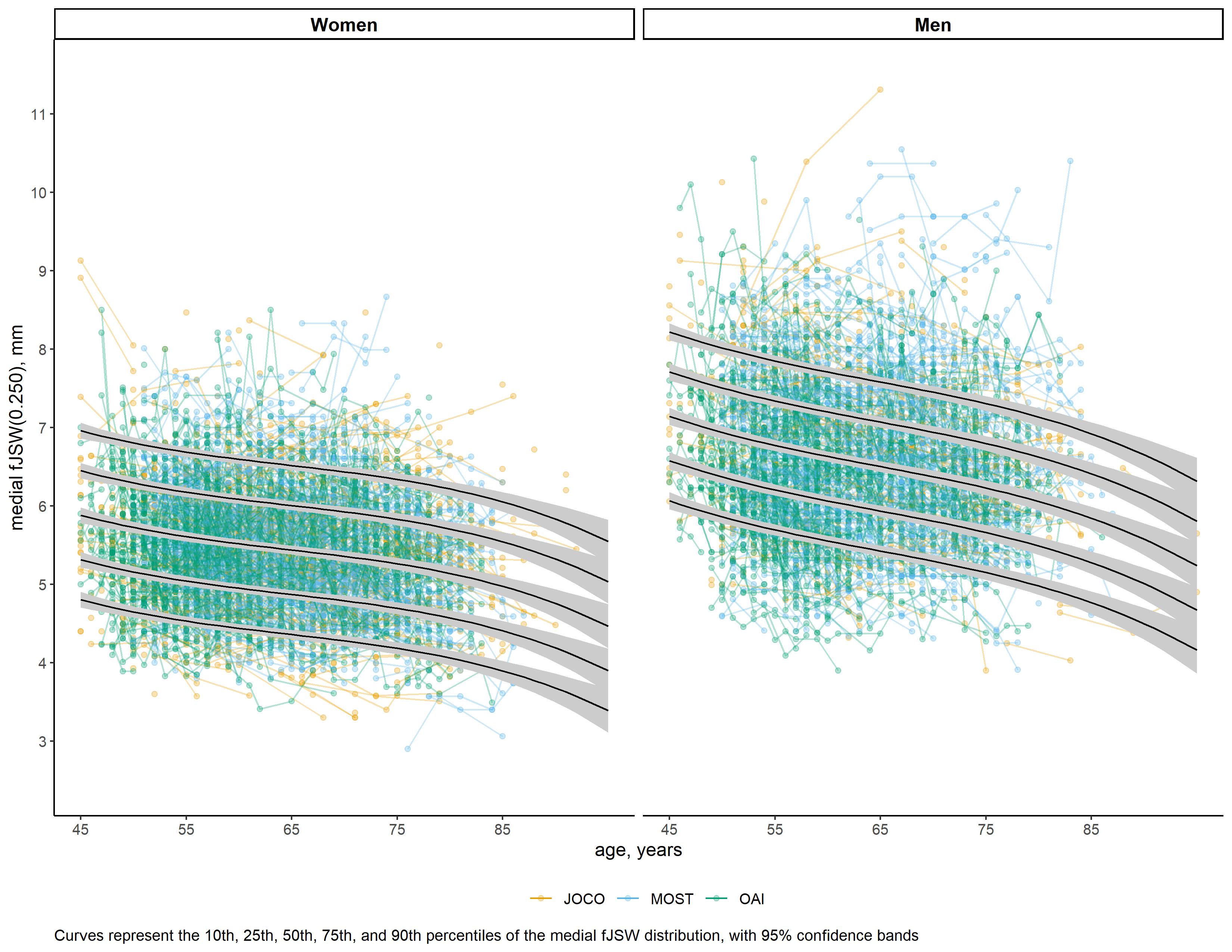
Results: The sample consisted of 3,116 participants who contributed 4,670 knees with at least one healthy time point, for a total of 12,347 healthy time point observations, including 2,349 from JoCoOA, 5,760 from MOST, and 4,238 from OAI. Participants self-identified as Non-Hispanic White (83%) and African American (17%), with 58% reporting female sex. Figure 1 shows sex-specific distributions of medial fJSW in healthy knees from age 45 to 90 years, with estimated percentile curves.
Conclusion: The natural history of medial fJSW among healthy knees is gradual loss with age. Women experiencing healthy knee aging had less fJSW than men across the spectrum of age. A large heterogeneity of medial fJSW in healthy knees was observed at all ages between 45 and 90 years. Healthy knee aging reference norms will aid our understanding of the natural history of fJSW.
Figure 1. Healthy knees by sex
K. Kwoh: Express Scripts, 2, Kolon Tissue Gene, 12, IDMC, Moebius, 2, Novartis, 1, Trial Spark, 2, Xalud, 1; E. Ashbeck: None; E. Bedrick: None; Y. Golightly: None; Z. Li: None; J. liew: None; A. Nelson: None; T. Neogi: None; X. Sun: None; J. Duryea: None.


.png "Kent Kwoh, MD photo")