University of Rochester School of Medicine and Dentistry Rochester, NY, United States
Disclosure information not submitted.
Kiana Chen1, Adelaide Weidner2, Olga Astapova2, Edward Schwarz2 and Homaira Rahimi2, 1University of Rochester School of Medicine and Dentistry, Rochester, NY, 2University of Rochester, Rochester, NY
Background/Purpose: Rheumatoid arthritis (RA) is characterized by chronic joint inflammation and bone erosion and is female predominant. The TNF-transgenic (TNF-Tg) murine model of RA develops inflammatory erosive arthritis and displays a sex difference in disease severity, with females having worse disease than males (1). Studies suggest androgens provide a protective effect against joint disease and TNF-mediated bone erosion (2). We have previously shown that the removal of endogenous sex hormones in TNF-Tg males significantly worsens their inflammatory erosive disease. Here, we investigated whether treatment of TNF-Tg mice with exogenous androgen ameliorates erosive disease.
Methods: TNF-Tg male mice were orchiectomized followed by subcutaneous implantation of either 5ɑ-dihydrotestosterone (DHT) or placebo pellet at 1-month old (n = 3/group). Pellets released 1.5mg of DHT or placebo for 60 days (0.025mg/day). Micro-computed tomography (µCT) scans of hindpaws were taken at 3-months old and compared with µCT data of same age intact TNF-Tg males (n = 4-6 paws/group). The total bone volumes (mm3) of the cuboid, talus, navicular and lateral intermediate cuneiform, and periarticular metatarsals were compared between groups. Deformation scores of the paws and weights were taken weekly from 1 to 3 months old. Same age sham TNF-Tg male mice weekly weights were compared between orchiectomized groups. Serum and paws were obtained for analysis and histology. Values are reported as the mean +/- standard deviation.
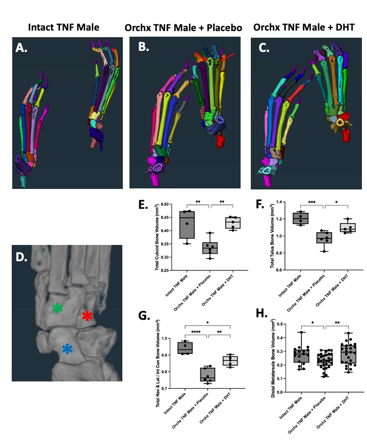
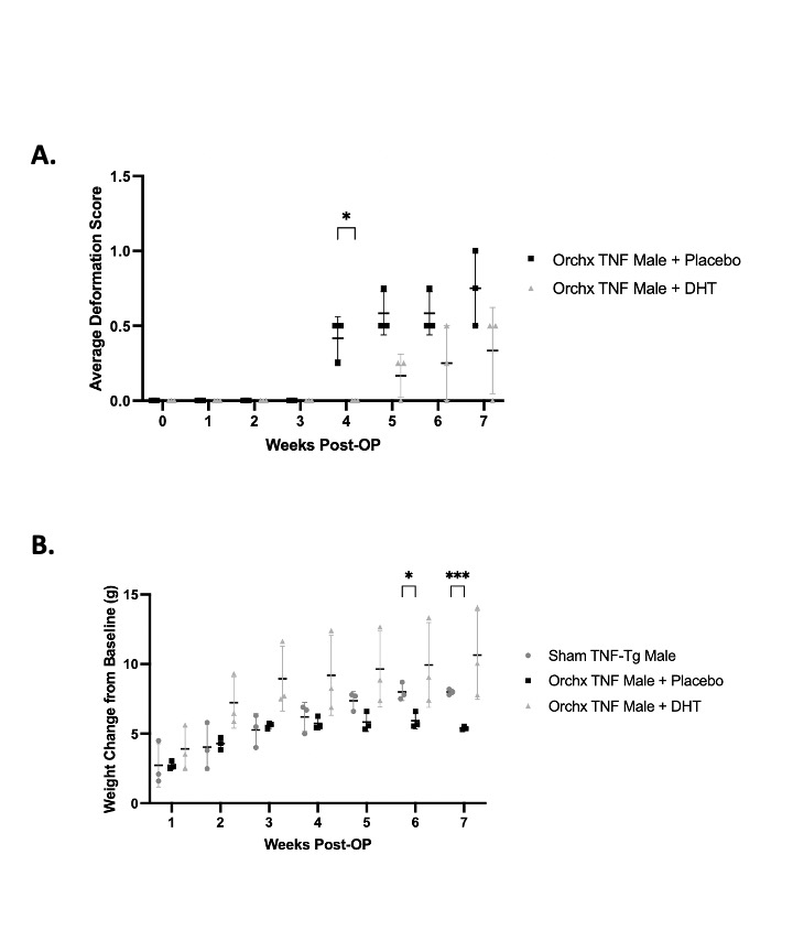
Results: Segmented hindpaw images showed bone erosion occurring in the periarticular regions of the metatarsals (Fig 1 A-C). Placebo-treated orchiectomized mice had significantly more bone volume loss than DHT-treated orchiectomized mice and intact mice in the cuboid (0.34 ± 0.03 Orchiectomized + Placebo; 0.43 ± 0.02 Orchiectomized + DHT; 0.43 ± 0.06 Intact), talus (0.97 ± 0.09 Orchiectomized + Placebo; 1.10 ±0.06 Orchiectomized + DHT; 1.21 ± 0.07 Intact), navicular and lateral intermediate cuneiform (0.78 ± 0.04 Orchiectomized + Placebo; 0.87 ± 0.03 Orchiectomized + DHT; 0.94 ± 0.04 Intact), and distal metatarsals (0.23 ± 0.05 Orchiectomized + Placebo; 0.28 ± 0.07 Orchiectomized + DHT; 0.27 ±0.07 Intact) (Fig 1D-H). Orchiectomized mice with placebo had significantly higher mean deformation scores at 4 weeks post-surgery (p = 0.03) (Fig 2A). Orchiectomized mice also gained significantly less weight than sham TNF-Tg mice by 6 weeks post-surgery (p = 0.03). DHT treatment of orchiectomized mice resolves that weight loss over time (Fig 2B).
Conclusion: Androgen treated orchiectomized arthritic mice had significantly improved bone volumes, limiting bone erosion even in the presence of ongoing inflammation. Clinical measures of weight loss and arthritis also improve with androgen treatment. These finding suggests sex hormones have a relationship with the immune system in inflammatory-erosive disease that warrants further study. Histological analysis of the paws and osteoclastogenic cultures of bone marrow are ongoing to delineate the mechanism of androgen effects on inflammation.
1. Bell R.D. et al. Arthritis Rheumatol 71(9): 1512-1523. 2019. 2. Traish A et al. J Clin Med 7(12): 549. 2018.
Figure 1. DHT Decreases Bone Erosions in Orchiectomized TNF-Tg Mice. µCT imaged hindpaw bones were segmented using Amira to determine total bone volume (mm3). Mice were divided into three groups (n = 4-6 paws/group), intact mice (A), orchiectomized (orchx) mice treated with placebo (B) and orchx mice treated with DHT (C). The cuboid (D, red asterisk), talus (D, blue asterisk), and navicular and lateral intermediate cuneiform (D, green asterisk) bone volumes were compared between groups. Orchiectomized mice treated with placebo were found to have significantly greater bone loss due to bone erosion in the cuboid (E), talus (F), navicular and lateral intermediate cuneiform (G) compared to the other cohorts. Placebo treated orchiectomized mice also had significantly less bone volume in the distal ends of the metatarsals that showed periarticular erosions reminiscent of erosions seen in RA patients (B, arrows; H). Orchiectomized mice treated with DHT had significantly greater bone volumes than orchiectomized mice treated with placebo, exhibiting that DHT treatment decreases bone erosion. Mid-hindpaw bone volume analysis was performed using a one-way ANOVA with Tukey’s multiple comparisons. Metatarsal bone volume analysis was performed using a one-way ANOVA with Fisher’s LSD test. * = p<0.05; ** = p<0.01; *** = p<0.001; **** = p<0.0001.
Figure 2. DHT-Treated Orchiectomized TNF-Tg Mice have Improved Clinical Measures of Disease. Orchiectomized mice treated with placebo and orchiectomized mice treated with DHT had weekly deformation scoring of their paws after surgery (n = 3 mice/cohort) (A). There was a significant difference in the average deformation score at week 4 post-op, signifying that orchiectomized mice treated with placebo displayed paw inflammation earlier than mice treated with DHT. Orchiectomized TNF-Tg mice treated with placebo, orchiectomized TNF-Tg mice treated with DHT, and sham TNF-Tg mice were also weighed weekly after surgery. The weight change from the baseline week (week 0) was compared between cohorts (B). Orchiectomized mice has significantly less weight gain than sham TNF-Tg mice by week 6 post-op. DHT treatment of orchiectomized mice ameliorates this weight loss over time. Average deformation score analysis was performed with multiple unpaired t-tests. Weight analysis was performed with a two-way ANOVA with Tukey’s multiple comparisons. * = p<0.05, *** = p<0.001.
K. Chen: None; A. Weidner: None; O. Astapova: None; E. Schwarz: None; H. Rahimi: None.