Hopital du Sacre-Coeur de Montreal Montréal, QC, Canada
Disclosure information not submitted.
Hussein Baalbaki1, David Dube1, Carolyn Ross2, Stephanie Ducharme-Benard3, Samer Hussein4, Rosalie-Selene Meunier4, Christian Pagnoux5 and Jean-Paul Makhzoum2, 1Hopital du Sacre-Coeur de Montreal, University of Montreal, Montreal, QC, Canada, 2Vasculitis Clinic, Canadian Network for Research on Vasculitides, Hopital du Sacre-Coeur de Montreal, Montreal, QC, Canada, 3Vasculitis Clinic, Hopital du Sacre-Coeur de Montreal, Montreal, QC, Canada, 4Vasculitis Clinic, Hopital du Sacre-Coeur de Montreal, Montreal, QC, Canada, 5Mount Sinai Hospital, Toronto, ON, Canada
Background/Purpose: Confirming the diagnosis of giant cell arteritis (GCA) remains challenging. Available diagnostic tests have limited accuracy and availability. Recently, optic nerve sheath enhancement on magnetic resonance imaging has been reported in patients with GCA. Whether this finding can be documented on ultrasound is unknown. Optic nerve (ON) ultrasound is non-invasive, easy to learn and does not require sophisticated equipment. This study aims to investigate if ON sheath measurement on ultrasound may be used to identify patients with active, new-onset GCA.
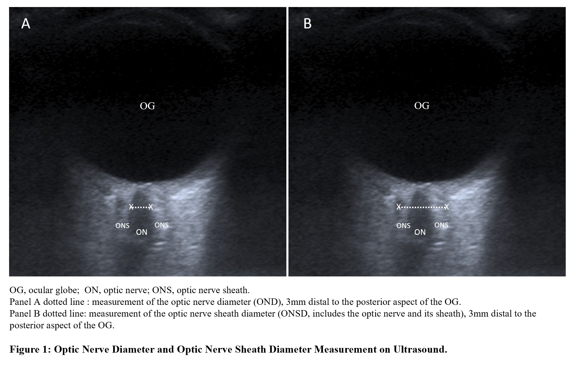
Methods: A cross sectional study was performed in our GCA Fast-Track clinic from June to December 2022. Adult patients were included if: (1) they were referred for suspected new-onset GCA, (2) had received < 14 days of glucocorticoids, and (3) had no prior history of intracranial hypertension, retinal disorder, or demyelinating disease. Study procedures were completed during the same visit: clinical assessment, bloodwork, ultrasound of temporal/axillary arteries and of the ON. Ultrasound measurements were performed for both eyes, 3mm distal to the posterior aspect of the ocular globe. Optic nerve sheath diameter (ONSD, includes ON and its sheath) and optic nerve diameter (OND) were measured. Optic nerve sheath thickness (ONST) was obtained by subtracting OND from ONSD (Figure 1). Final diagnosis of GCA was confirmed clinically after 6-month follow-up. Independent sample t-test was used to compare mean differences in ONSD, OND and ONST in patients with and without GCA at baseline. Paired-sample t-test was used to compare measurements between both eyes. Multivariable analysis was conducted for clinical baseline characteristics and ON ultrasound measurements. A significance level of 0.05 with Bonferroni correction (n=6) for multiple comparisons was used.
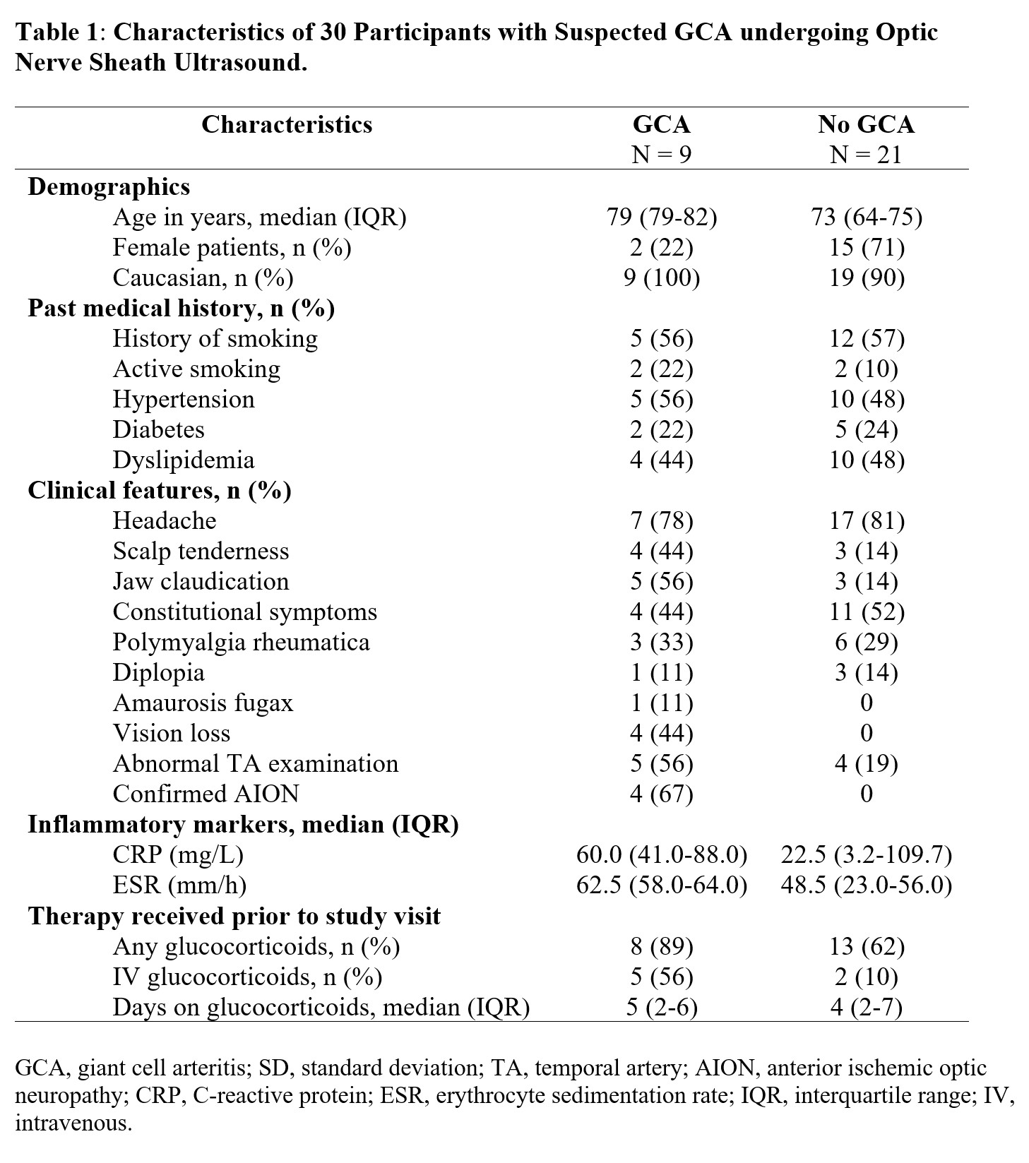
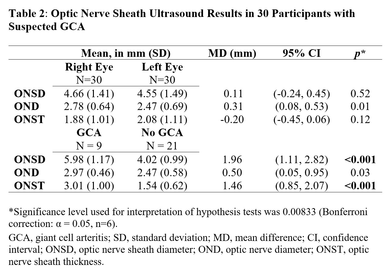
Results: A total of 30 participants were enrolled, including 9 participants with a final diagnosis of GCA (all of whom also satisfied 2022 ACR/EULAR classification criteria (Table 1)). Patients with GCA had a higher mean ONSD (5.98mm) compared to those without GCA (4.02mm); with a mean difference of 1.96mm (95% confidence interval [CI]: 1.11–2.82, p< 0.001). Mean ONST was higher in patients with GCA (3.01mm) than those without GCA (1.54mm), with a mean difference of 1.46mm (95%CI: 0.85–2.07, p< 0.001). Mean OND was numerically but not statistically higher in patients with GCA (Table 2). Paired measurement comparisons of ONSD, OND, ONST did not differ between the right and left eyes. On multivariable analysis, there was no significant association between age, sex, hypertension, diabetes, anterior ischemic optic neuropathy and ONSD, OND, ONST ultrasound measurements.
Conclusion: Patients with GCA had significantly wider optic nerve sheaths on ultrasound than patients without GCA. ON ultrasound may represent a novel, rapid, bedside diagnostic test in GCA. A large prospective cohort study is ongoing to confirm these findings and evaluate if ONSD-ONST are dynamic disease markers in GCA (ClinicalTrials.gov id: NCT05749094).
H. Baalbaki: None; D. Dube: None; C. Ross: None; S. Ducharme-Benard: None; S. Hussein: None; R. Meunier: None; C. Pagnoux: AstraZeneca, 1, 2, 6, GlaxoSmithKlein(GSK), 1, 6, Otsuka, 1, 2, 5, 6, Pfizer, 5, Roche, 2; J. Makhzoum: None.


.jpg "Hussein Baalbaki, MD photo")