Session: (1383–1411) Spondyloarthritis Including Psoriatic Arthritis – Diagnosis, Manifestations, & Outcomes Poster II: Imaging & AS
1396: Comprehensive Evaluation of the Digital Annular Pulleys: From Ultrasonographical to Anatomical and Histological Evaluation with Special Focus on the Entheses
Luis Coronel1, Peter Mandl2, María Isabel Miguel3, Joan Blasi4, Maria Lopez-Lasanta1, Sara Marsal1, Maria-Antonietta D'Agostino5 and Ingrid Moller6, 1Hospital Universitari Vall d'Hebron, Rheumatology, Barcelona, Spain, 2Department of Rheumatology, Medical University of Vienna, Vienna, Austria, 3Universitat de Barcelona - Campus Bellvitge, Unitat d’anatomía i embriología humana, L'Hospitalet de Llobregat, Barcelona, Spain, 4Universitat de Barcelona - Campus Bellvitge, Histologia, L'Hospitalet de Llobregat, Barcelona, Spain, 5Universita Cattolica del Sacro Cuore Rome, Courbevoie, France, 6Instituto Poal de Reumatología, Barcelona, Spain
Background/Purpose: The digital annular pulleys (DAP) are anatomical structures of soft connective tissue organized as transverse fibers of variable width, thickness, and configuration that overlay the synovial sheath of the flexor tendons. These structures prevent the flexor tendons from bowing and maintain them in constant relationship with the joint axis of motion. The thickening of DAP has been described as one of the elementary lesions involved in dactylitis in psoriatic arthritis (PsA) patients highlighting the importance of functional pulleys in the pathophysiological manifestations of PsA, as a form of functional enthesis. The present study assesses the ultrasonographic, anatomical and histological characteristics of the DAP anatomical entheses.
Methods: DAP from 20 formalin-embalmed cadaveric hands were assessed with grey-scale ultrasound (US).The US evaluations, performed by 2 expert sonographers, included identification, width, and characterization of insertion sites of the annular pulleys with a newly proposed dynamic maneuver that grants a better exposure of DAP entheses (Figure 1). Then, dissection was performed from the midcarpal area to the distal end of each finger, second to fifth (Figure 2). Measurements of the thickness of the DAP were obtained with a digital caliper. Intra and inter-reader reliability was evaluated using Cohen´s Kappa for all the evaluations. For the histological study, the samples were decalcified using EDTA, sectioned by microtome, and fixed in buffered formalin for two days and, stained with hematoxylin-eosin. The slides were observed and measured with a Leica DMD 108 microscope. Quantitative demographic variables were summarized in means and standard deviations (SD) and qualitative variables in frequencies and percentages (n [%]). Quantitative and qualitative demographic variables were compared across groups using Student's t-test and chi-square and Fisher test, respectively.All tests were two-sided and p-values < 0.05 were considered statistically significant. Statistical analyses were performed using R studio (program version RStudio 2022.12.0+353).
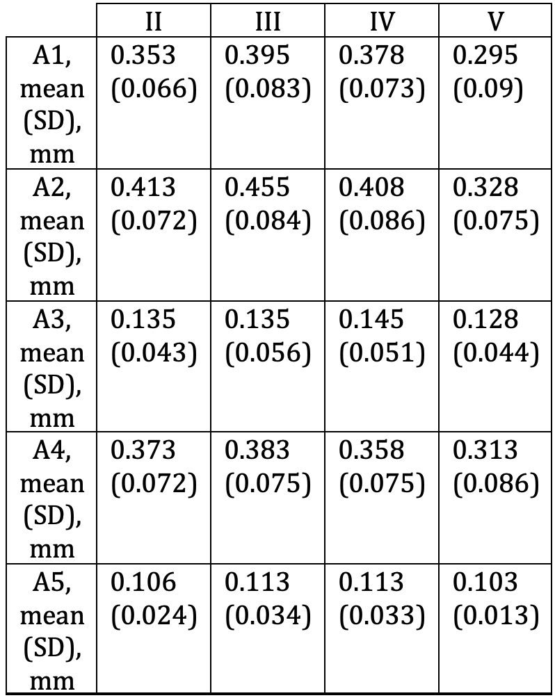
Results: The mean age of donors and HC was 81±9 years; range 58–96 years and 11 were females (55%). The mean values of DAP thickness detected by US were analyzed for each finger. Detailed results are presented in Table 1. The intra-reader (mean 0.85, Standard Deviation 0.11) and inter-reader reliability (mean: 0.82, 95% confidence interval: 0.81-0.82) was high. A high correlation between ultrasonographic and anatomical measurements was found (r=0.96). The histological description of the DAP shows a well-vascularized outer surface and a fibrocartilaginous component in the inner surface. The insertion points at the sesamoid or the phalangeal bones demonstrate a diverse nature of the enthesis with fibrocartilaginous and fibrous areas.
Conclusion: These preliminary results show that DAP have a mixed fibrous and fibrocartilaginous enthesis and US is a valuable tool for identifying DAPs' width and anatomical structures, with good correlation with the histological study.
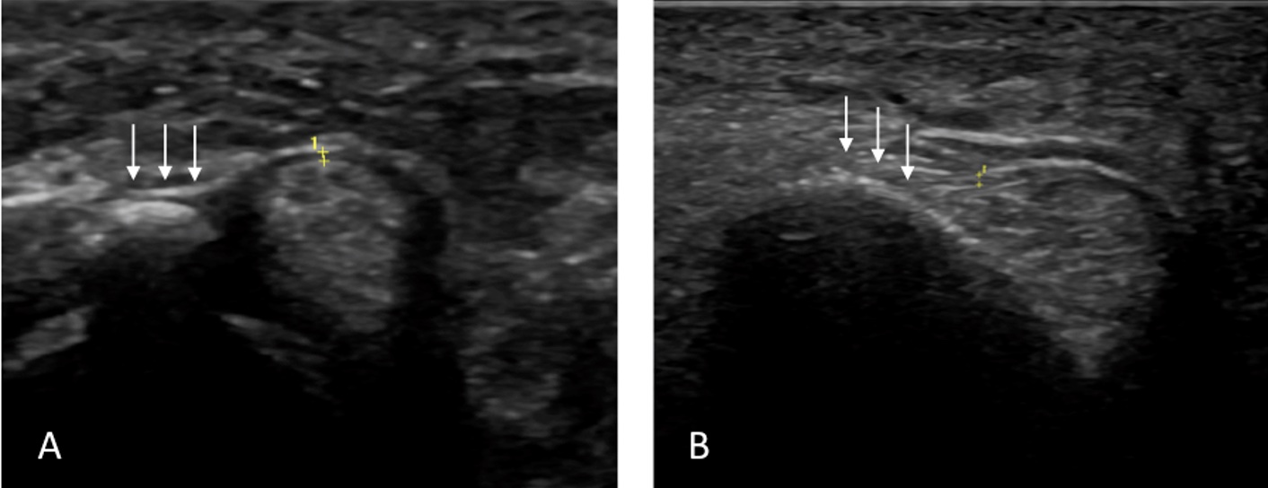
Figure 1. Ultrasound longitudinal scans of the annular pulleys entheses. A) A1 enthesis at the sesamoid bone. B) A2 enthesis in the proximal phalanx. White arrows: Annular pulley entheses. Yellow stars: Dorsal and volar limits of the pulley.
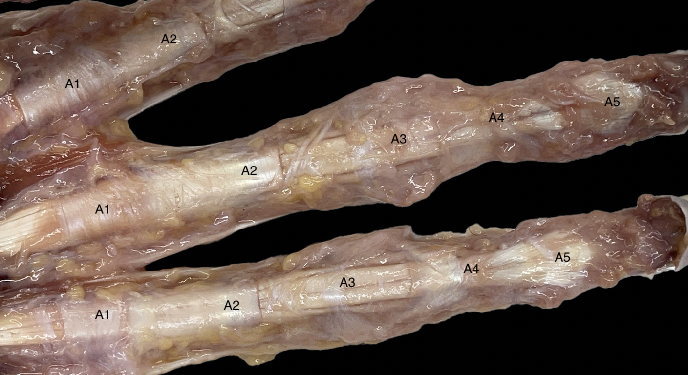
Figure 2. Anatomical dissection of the fingers exposing DAP from A1 to A5.
Table 1. Mean values of pulleys thickness for all annular pulleys (A1-A5) in fingers II-V.
L. Coronel: None; P. Mandl: AbbVie/Abbott, 5, 6, Bristol-Myers Squibb(BMS), 6, Celgene, 6, Eli Lilly, 6, Janssen, 6, Novartis, 5, 6, Pfizer, 5, Roche, 6, UCB, 5, 6; M. Miguel: None; J. Blasi: None; M. Lopez-Lasanta: None; S. Marsal: None; M. D'Agostino: None; I. Moller: None.