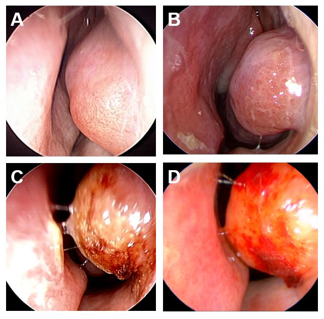
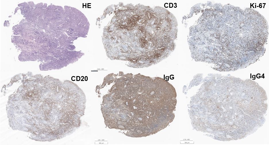
Seoul National University Hospital Seoul, South Korea
Disclosure information not submitted.
Ji In Jung1, Ju Yeon Kim2, Jun Won Park2, Eun Bong Lee2, Jin Kyun Park3 and Doo Hee Han2, 1Division of Rheumatology, Department of Internal Medicine, Seoul National University Hospital, Seoul, South Korea, 2Seoul National University Hospital, Seoul, South Korea, 3Seoul National University College of Medicine, Seoul, South Korea
Background/Purpose: IgG4-related disease (IgG4-RD) is a rare, multi-system, fibro-inflammatory disorder characterized by the infiltration of lymphoplasmacytic cells, storiform fibrosis, obliterative phlebitis, and the presence of IgG4-positive plasma cells in affected tissues, accompanied by elevated serum IgG4 levels. In IgG4-RD, sinonasal involvement presents as nasal obstruction, chronic sinusitis, and bleeding. Nasal obstruction may arise from a sessile submucosal mass located in the anterior portion of the inferior turbinate, exhibiting dense lymphocytic lymphoplasmacytic infiltration. We aimed to investigate whether these lymphoid masses in the nasal inferior turbinate represent a nasal manifestation of IgG4-RD or a distinct chronic inflammatory disease entity that has yet to be defined.
Methods: This retrospective study enrolled patients who underwent endoscopy at Seoul National University Hospital between January 2020 and May 2023 due to nasal symptoms, including nasal obstruction or nasal crust. The clinical characteristics and laboratory parameters, such as erythrocyte sedimentation rate (ESR), high-sensitivity C-reactive protein (hs-CRP), and serum IgG4 levels, were evaluated (Figure 1). Biopsies were performed on nasal lymphoid masses exhibiting damaged mucosa features like crusting and bleeding. The diagnosis of IgG4-RD was based on the 2019 classification criteria of IgG4-RD from the American College of Rheumatology/European League Against Rheumatism. All data was analyzed using Chi-square test and Student's t-test.
Results: In this study, a total of 14103 patients were evaluated for nasal symptoms, and among them, 39 patients presented with lesions resembling lymphoid follicles in the inferior turbinate upon endoscopic evaluation (Figure 2). Among the 14 patients who underwent biopsy, all of them exhibited dense lymphoplasmacytic infiltrates and more than 10 IgG4-expressing plasma cells per high-power field (HPF) (Figure 3). Among these patients, 7 out of 14 (50%) had an IgG4+/IgG+ ratio exceeding 40% and met the histologic criteria for IgG4-RD. However, only 1 patient had elevated serum IgG4 levels. None of the patients were classified as having IgG4-RD based on the EULAR/ACR classification criteria. Patients with significant nasal symptoms who received surgical resection experienced improvement without recurrence of local symptoms.
Conclusion: To the best of our knowledge, this study provides the first comprehensive description of a nasal sessile lymphoid mass located in the inferior turbinate. Our findings indicate that this lymphoid mass, characterized by IgG4+ lymphoplasmacytic infiltration, is likely to represent a distinct disease entity separate from the sinonasal involvement of IgG4-RD. Further study is required to define the optimal treatment and long prognosis.
Figure 1. Patient flow
Figure 2. Representative endoscopic findings lymphoid mass. Rhinoscopy reveals yellowish sessile submucosal mass with intact overlying mucosa in the anterior portion of the inferior turbinate - (A,B) Mild and (C,D) severe cases. (D) Overlying mucosa was covered with bloody crust.
Figure 3. Histology of nasal polyp
J. Jung: None; J. Kim: None; J. Park: None; E. Lee: Pfizer, 2; J. Park: None; D. Han: None.