Department of Rheumatology, Nanfang Hospital, Southern Medical University Guangzhou, Guangdong, China
Disclosure information not submitted.
Yi Feng, Jiayu Qin, Lijuan Zheng, Hao Ren, Min Yang and Qin Huang, Department of Rheumatology, Nanfang Hospital, Southern Medical University, Guangzhou, China
Background/Purpose: Cognitive dysfunction is one of the most common manifestations of neuropsychiatric systemic lupus erythematosus (NPSLE) and severely affects patients' health-related quality of life. Intermittent fasting (IF) has been shown promising therapeutic effects in neurodegenerative diseases. In this study, we evaluated the impact of intermittent fasting on cognitive function in lupus-prone mice.
Methods: MRL/lpr mice, as an animal model for studying NPSLE, can spontaneously exhibit cognitive dysfunction. After 8 weeks of alternate-day fasting, novel object recognition and Morris water maze tests were used to assess cognitive manifestation. The number of microglia (IBA-1+) was evaluated by immunofluorescence staining. Expression levels of IL-1β, IL-6 and TNF-α were detected by qRT-PCR. The blood-brain barrier permeability and autophagy levels were evaluated by western blotting.
Results: We found that IF improves cognitive function of MRL/lpr mice in the behavioral tests. IF also decreased hippocampal microglia activation and expression of inflammatory cytokines in MRL/lpr mice. In addition, we demonstrated that IF reduced blood-brain barrier permeability. Furthermore, IF inhibited mTOR signaling and increased autophagy levels.
Conclusion: These data indicate that IF improves cognitive function in lupus-prone mice.
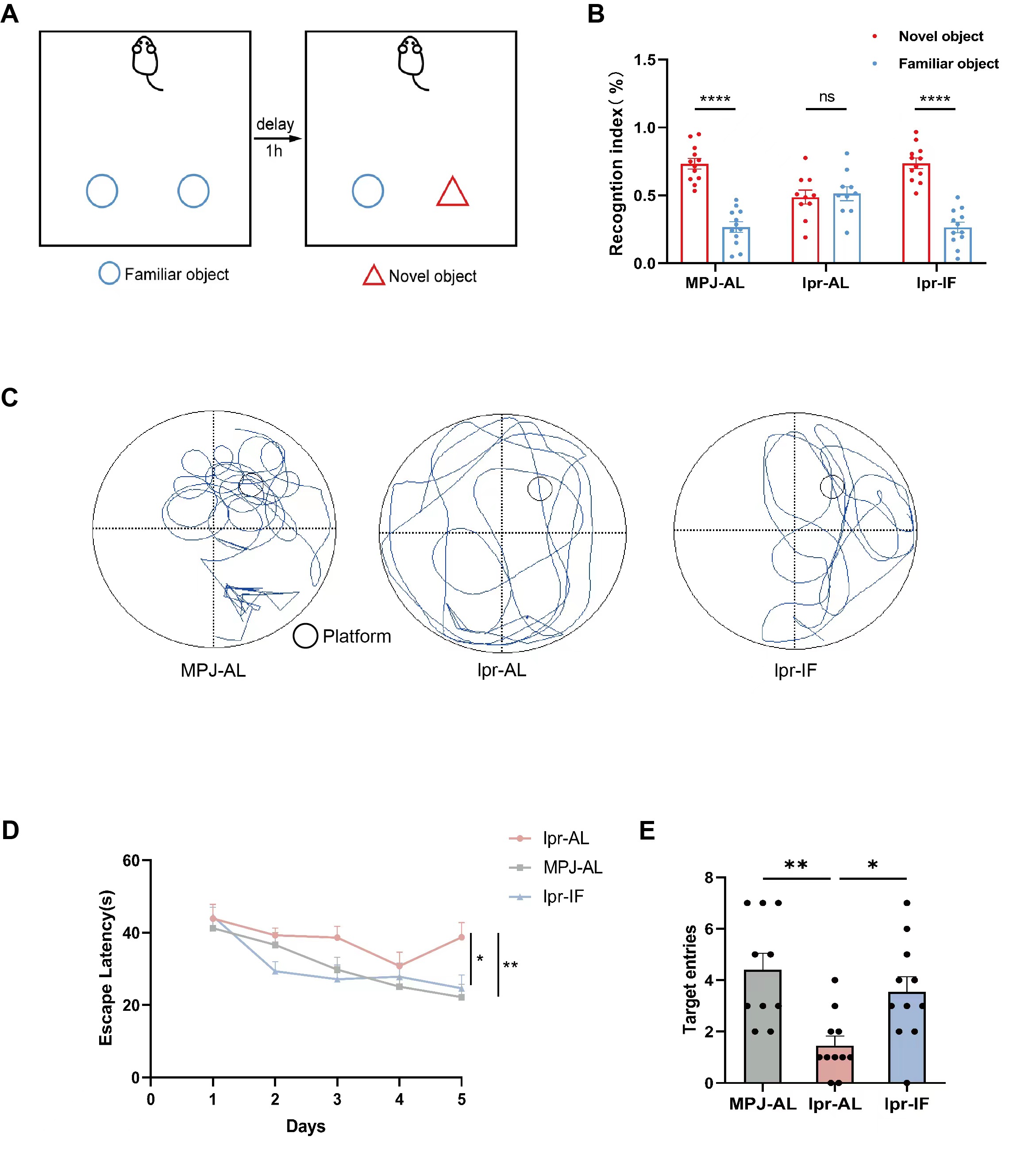
Fig.1 IF improves cognitive function of MRL/lpr mice.A:The experimental procedure of the novel object recognition test.B: The test recognition index of mice (n = 10-12 mice per group).C:The represent image of day6 in the Morris water maze test.D:The escape latency to the platform of day1-5.E:The target entries of day6.The data are represented as the mean ± standard error of the mean. *P < 0.05, **P < 0.01, ***P < 0.001 and ****P < 0.0001; NS, not significant.
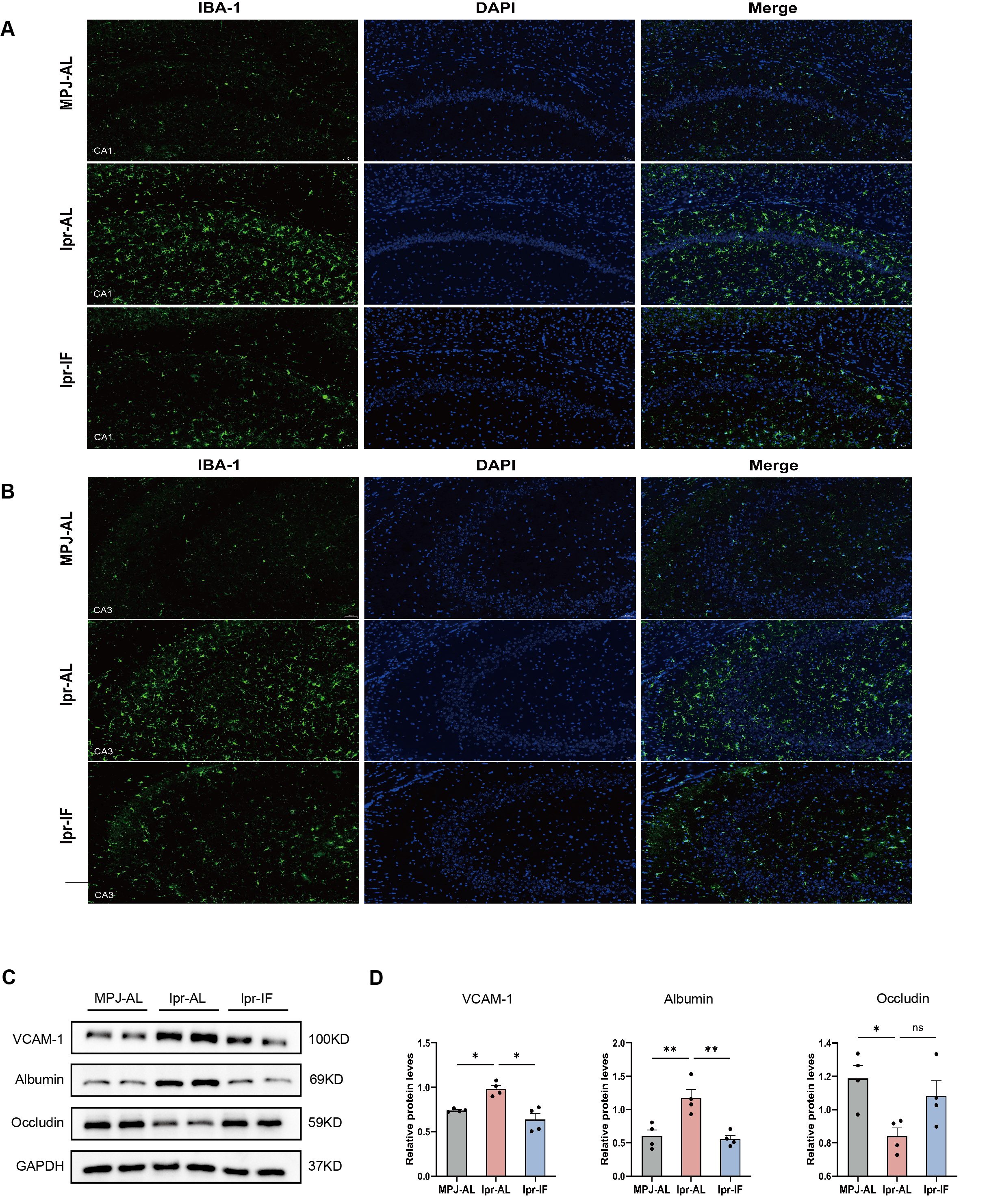
Fig.2 IF decreased hippocampal microglia activation and blood-brain barrier permeability of MRL/lpr mice.A-B:The represent image of microglia (IBA-1+) in the CA1 and CA3 of hippocampal.C:Western blotting analysis of proteins in the brain of mice.D:The quantification of indicated protein levels shown in A (n = 4 mice per group).
Fig.3 IF inhibited mTOR signaling and increased autophagy levels of MRL/lpr mice.A:Western blotting analysis of proteins in the hippocampal of mice.B:The quantification of indicated protein levels shown in A (n = 4 mice per group).
Y. Feng: None; J. Qin: None; L. Zheng: None; H. Ren: None; M. Yang: None; Q. Huang: None.