0813: Sonographic Crystal Deposits and Power-Doppler Signal in Patients with Gout Fulfilling Remission Criteria: A Multicenter Study Enrolling 115 Participants
Dr Balmis Alicante General University Hospital-ISABIAL Alicante, Spain
Disclosure information not submitted.
Mariano Andrés1, Nalia Domínguez-Lirón2, Enrique Calvo-Aranda3, Esther Vicente Rabaneda4, Agustín Martínez-Sanchís1, Francisca Sivera5, Diana Peiteado6, Alejandro Prada7, Blanca Garcia8, Basilio Rodríguez9, BORIS ANTHONY BLANCO CACERES10, José Antonio Bernal11, Santos Castañeda12, Laura Barrio7, Sonia Minguez9, Mónica Vázquez Díaz13, José Miguel Senabre11, Cristina Bohorquez14, Silvia Gómez-Sabater15, Rocío Caño-Alameda15 and Eugenio De Miguel16, 1Dr Balmis Alicante General University Hospital-ISABIAL, Alicante, Spain, 2Miguel Hernández University, Alicante, Spain, 3Hospital Universitario Infanta Leonor, Madrid, Spain, 4Rheumatology, Hospital Universitario de La Princesa, Madrid, Spain, 5Elda General University Hospital, Elda, Spain, 6Hospital La Paz, Madrid, Spain, 7Hospital Universitario de Torrejón, Madrid, Spain, 8Hospital Universitario Puerta de Hierro Majadahonda, Madrid, Spain, 9Fundació Althaia, Manresa, Spain, 10Hospital Universitario Ramon y Cajal, Madrid, Spain, 11Hospital Marina Baixa, Villajoyosa, Spain, 12Hospital Universitario de la Princesa, Madrid, Spain, 13Hospital Ramon y Cajal, Madrid, Spain, 14Rheumatology Unit, Hospital Universitario Príncipe de Asturias, Alcalá de Henares, Spain, 15Rheumatology Department, Dr. Balmis University General Hospital, Alicante. Institute for Health and Biomedical Research (ISABIAL), Alicante, Spain, 16Hospital Universitario La Paz, Madrid, Spain
Background/Purpose: The prevalence of sonographic monosodium urate (MSU) crystal deposition and inflammation in patients with gout in remission is unknown. In 2022, we reported a preliminary estimation of 88.7% of patients with persistent deposits, while one out of three had a positive power-Doppler (PD) signal [1]. This abstract communicates the sonographic evaluation of our initial 115 patients with gout in remission.
Methods: Observational cross-sectional multicenter study. Consecutive patients with gout (ACR/EULAR classification criteria +/- MSU crystal-proven) who met preliminary remission criteria [2] were recruited at eleven Spanish rheumatology units. They underwent a sonographic scanning of the first metatarsophalangeal and second metacarpophalangeal joints, knees, talar cartilages, and patellar and Achilles tendons. The sonographers were blinded to participants' clinical and laboratory data. We determined the prevalence (with 95% confidence interval -CI) of sonographic MSU crystal deposits (tophi, aggregates, and double contour sign) and inflammation by power Doppler [PD] signal (graded as 0-3, positive if ≥1). Associations between deposits and PD signal and clinical and laboratory variables were also analyzed by chi-2 and logistic regression.
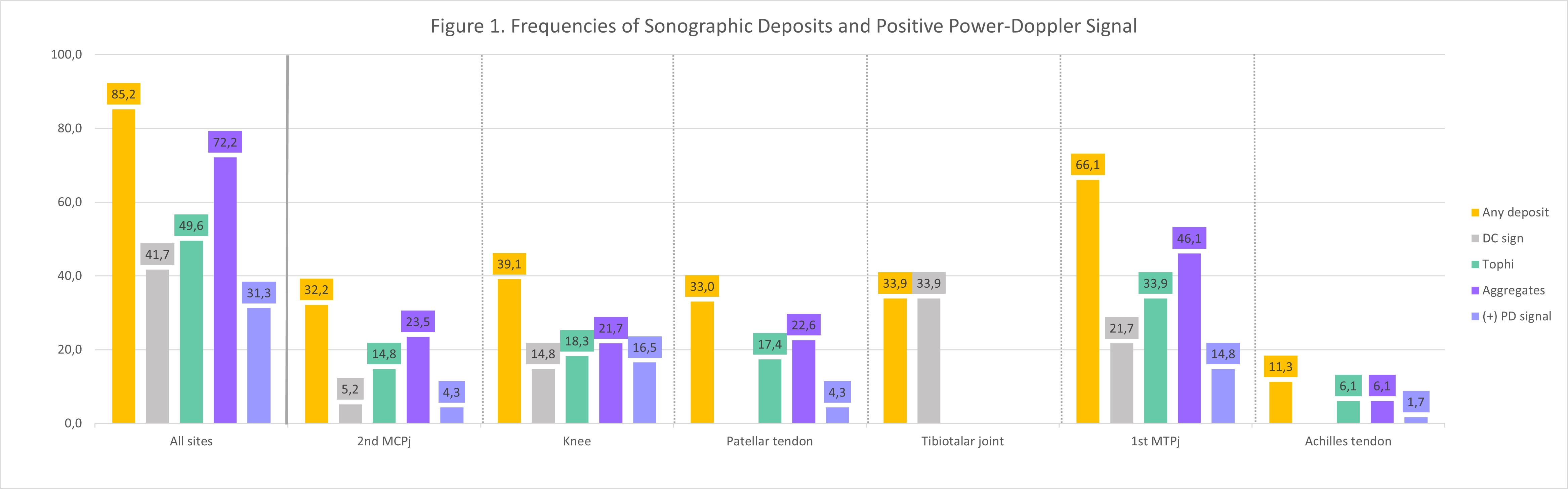
Results: The sample includes 115 participants, mean age of 65.2 years (SD 9.7), 93.9% males. The mean gout duration was 13.9 years (SD 10.9), and the disease was tophaceous at baseline in 15.7%. The mean serum urate level in the preceding year was 4.5 mg/dl (SD 0.9), with a mean duration of urate-lowering therapy of 55.8 months (SD 34.7). The prevalence of deposits in at least one location was 85.2% (95%CI 77.6-90.6%), with a median of 3 locations with deposits (range 0-9). Articular deposits (80.0%) were more common than tendinous deposits (39.1%), and aggregates were the most frequent sonographic finding (83.0%) [Figure 1]. If aggregates are not considered, prevalent deposits dropped to 73.0% (95%CI 64.3-80.3%). A positive PD signal was present in 31.3% of participants (95%CI 23.6-40.3%), mainly at joints (27.8%). Rates of deposits and positive PD signals were mildly lowered when restricted to four locations (82.6% and 27.8%, respectively) and any site except 1MTP joints (77.4% and 22.6%). A significant association between deposits and positive PD signal was confirmed at joints (p=0.005) and tendons (p=0.033). No secondary variable was associated with deposits or positive PD signal.
Conclusion: Our updated multicenter dataset confirms that most patients with gout fulfilling remission criteria still show sonographic MSU crystal deposits and one third, sonographic inflammation. The relevance of persistent deposits and inflammation in this setting needs further clarification.
References: [1] Domínguez-Lirón N. Arthritis Rheumatol 2022;74(suppl 9). [2] de Lautour H. Arthritis Care Res 2016;68(5):667.
Figure 1.
M. Andrés: None; N. Domínguez-Lirón: None; E. Calvo-Aranda: None; E. Vicente Rabaneda: None; A. Martínez-Sanchís: None; F. Sivera: AbbVie/Abbott, 1, AstraZeneca, 5, Bristol-Myers Squibb(BMS), 5, Eli Lilly, 5, GlaxoSmithKlein(GSK), 6, Novartis, 5, 6, Pfizer, 1, Roche, 5, UCB, 6; D. Peiteado: None; A. Prada: None; B. Garcia: None; B. Rodríguez: None; B. BLANCO CACERES: None; J. Bernal: None; S. Castañeda: None; L. Barrio: None; S. Minguez: None; M. Vázquez Díaz: None; J. Senabre: None; C. Bohorquez: None; S. Gómez-Sabater: None; R. Caño-Alameda: None; E. De Miguel: None.